Shunsuke Nakamura, Hiroya Shimauchi-Ohtaki, Fumiaki Honda, Yuta Goto, Satoshi Nakata, Keishi Horiguchi, Ran Tomomasa, Junko Hirato, Hideaki Yokoo, Soichi Oya
{"title":"A Primary Spinal Intramedullary Mixed Germ Cell Tumor: A Case Report and Literature Review.","authors":"Shunsuke Nakamura, Hiroya Shimauchi-Ohtaki, Fumiaki Honda, Yuta Goto, Satoshi Nakata, Keishi Horiguchi, Ran Tomomasa, Junko Hirato, Hideaki Yokoo, Soichi Oya","doi":"10.2176/jns-nmc.2024-0202","DOIUrl":null,"url":null,"abstract":"<p><p>Central nervous system germ cell tumors are rare and account for 2% to 3% of all central nervous system tumors in Japan. Here, we report an extremely rare case of a primary spinal intramedullary mixed germ cell tumor. A 33-year-old man presented with a chief report of dysuria and numbness in the right lower extremity. Magnetic resonance imaging revealed a mass lesion on the left side of the intramedullary spinal cord at the Th10-11 vertebral body level with hyperintensity on T2-weighted images, isointensity on T1-weighted images, and uniform contrast in gadolinium. Cerebrospinal fluid examination revealed a few atypical cells. Although tumor removal using the posterior median sulcus approach was attempted, only a biopsy was performed because intraoperative rapid pathology suggested a possible diagnosis of germinoma. Permanent pathology revealed a mixed germ cell tumor (mainly comprising a germinoma with a yolk sac tumor). Postoperatively, cerebrospinal irradiation and 8 courses of the carboplatin and etoposide regimen were administered. No recurrence or new lesions were observed on magnetic resonance imaging at 94 months postoperatively. Our extensive literature search found only 4 cases of a mixed germ cell tumor of primary intramedullary origin in the spinal cord. Most spinal germ cell tumors described in the literature are either germinomas or mature teratomas; however, mixed germ cell tumors can also occur, albeit infrequently. Although additional cases need to be accumulated, our case suggests that yolk sac elements in spinal mixed germ cell tumors might not be directly associated with poor life expectancy.</p>","PeriodicalId":101331,"journal":{"name":"NMC case report journal","volume":"12 ","pages":"53-58"},"PeriodicalIF":0.0000,"publicationDate":"2025-02-07","publicationTypes":"Journal Article","fieldsOfStudy":null,"isOpenAccess":false,"openAccessPdf":"https://www.ncbi.nlm.nih.gov/pmc/articles/PMC11867697/pdf/","citationCount":"0","resultStr":null,"platform":"Semanticscholar","paperid":null,"PeriodicalName":"NMC case report journal","FirstCategoryId":"1085","ListUrlMain":"https://doi.org/10.2176/jns-nmc.2024-0202","RegionNum":0,"RegionCategory":null,"ArticlePicture":[],"TitleCN":null,"AbstractTextCN":null,"PMCID":null,"EPubDate":"2025/1/1 0:00:00","PubModel":"eCollection","JCR":"","JCRName":"","Score":null,"Total":0}
引用次数: 0
Abstract
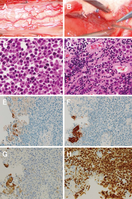
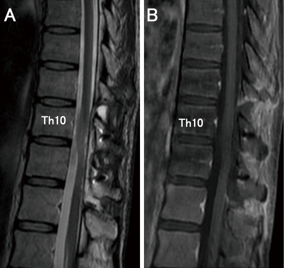
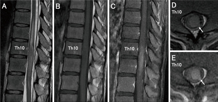
Central nervous system germ cell tumors are rare and account for 2% to 3% of all central nervous system tumors in Japan. Here, we report an extremely rare case of a primary spinal intramedullary mixed germ cell tumor. A 33-year-old man presented with a chief report of dysuria and numbness in the right lower extremity. Magnetic resonance imaging revealed a mass lesion on the left side of the intramedullary spinal cord at the Th10-11 vertebral body level with hyperintensity on T2-weighted images, isointensity on T1-weighted images, and uniform contrast in gadolinium. Cerebrospinal fluid examination revealed a few atypical cells. Although tumor removal using the posterior median sulcus approach was attempted, only a biopsy was performed because intraoperative rapid pathology suggested a possible diagnosis of germinoma. Permanent pathology revealed a mixed germ cell tumor (mainly comprising a germinoma with a yolk sac tumor). Postoperatively, cerebrospinal irradiation and 8 courses of the carboplatin and etoposide regimen were administered. No recurrence or new lesions were observed on magnetic resonance imaging at 94 months postoperatively. Our extensive literature search found only 4 cases of a mixed germ cell tumor of primary intramedullary origin in the spinal cord. Most spinal germ cell tumors described in the literature are either germinomas or mature teratomas; however, mixed germ cell tumors can also occur, albeit infrequently. Although additional cases need to be accumulated, our case suggests that yolk sac elements in spinal mixed germ cell tumors might not be directly associated with poor life expectancy.


 分享
分享

 求助内容:
求助内容: 应助结果提醒方式:
 扫码关注我们
