{"title":"The three-dimensional finite element model of unilateral complete cleft lip and palate and mechanical analysis of the oral surfaces.","authors":"Qingqian Wei, Hao Liang, Jingyi Wang, Fei Chen, Yinyue Chen, Yiwei Liu, Haidong Li","doi":"10.1186/s40902-024-00452-7","DOIUrl":null,"url":null,"abstract":"<p><strong>Background: </strong>Cleft palate is a prevalent oral and maxillofacial malformation that requires complex surgical interventions. In cleft palate repair, managing flap tension is critical to avoid complications such as flap rupture and impaired healing. Additionally, excessive flap movement can compromise blood supply, affecting postoperative outcomes. A thorough understanding of these biomechanical factors is crucial for surgical success.</p><p><strong>Methods: </strong>A three-dimensional finite element model was developed using CT scan data to simulate the biomechanical behavior of the cleft palate under surgical conditions. The model was constructed and analyzed using ANSYS Workbench and related software, incorporating material properties of bone, mucosa, and muscle. Stress and deformation distributions were calculated to evaluate surgical incision points and flap movement.</p><p><strong>Results: </strong>The model identified critical areas of high tension and movement along the surgical incisions on both oral and nasal surfaces. The maximum deformation observed was 3.9885 mm, with stress concentration points along the suture lines and flap edges. The results highlighted specific regions prone to mechanical stress, which are crucial for optimizing surgical strategies.</p><p><strong>Conclusion: </strong>This study demonstrates the potential of a 3D finite element model in predicting mechanical responses of the cleft palate during surgical repair. The findings provide surgeons with valuable insights for improving incision placement, flap design, and suturing techniques to minimize tension and enhance healing. This personalized approach could significantly improve surgical outcomes and reduce postoperative complications in cleft palate repair.</p>","PeriodicalId":18357,"journal":{"name":"Maxillofacial Plastic and Reconstructive Surgery","volume":"47 1","pages":"6"},"PeriodicalIF":2.8000,"publicationDate":"2025-03-05","publicationTypes":"Journal Article","fieldsOfStudy":null,"isOpenAccess":false,"openAccessPdf":"https://www.ncbi.nlm.nih.gov/pmc/articles/PMC11883061/pdf/","citationCount":"0","resultStr":null,"platform":"Semanticscholar","paperid":null,"PeriodicalName":"Maxillofacial Plastic and Reconstructive Surgery","FirstCategoryId":"1085","ListUrlMain":"https://doi.org/10.1186/s40902-024-00452-7","RegionNum":0,"RegionCategory":null,"ArticlePicture":[],"TitleCN":null,"AbstractTextCN":null,"PMCID":null,"EPubDate":"","PubModel":"","JCR":"Q2","JCRName":"DENTISTRY, ORAL SURGERY & MEDICINE","Score":null,"Total":0}
引用次数: 0
Abstract
Background: Cleft palate is a prevalent oral and maxillofacial malformation that requires complex surgical interventions. In cleft palate repair, managing flap tension is critical to avoid complications such as flap rupture and impaired healing. Additionally, excessive flap movement can compromise blood supply, affecting postoperative outcomes. A thorough understanding of these biomechanical factors is crucial for surgical success.
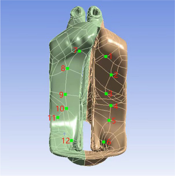
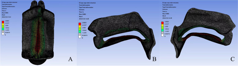
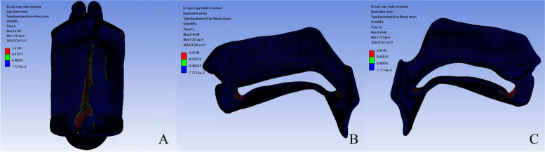
Methods: A three-dimensional finite element model was developed using CT scan data to simulate the biomechanical behavior of the cleft palate under surgical conditions. The model was constructed and analyzed using ANSYS Workbench and related software, incorporating material properties of bone, mucosa, and muscle. Stress and deformation distributions were calculated to evaluate surgical incision points and flap movement.
Results: The model identified critical areas of high tension and movement along the surgical incisions on both oral and nasal surfaces. The maximum deformation observed was 3.9885 mm, with stress concentration points along the suture lines and flap edges. The results highlighted specific regions prone to mechanical stress, which are crucial for optimizing surgical strategies.
Conclusion: This study demonstrates the potential of a 3D finite element model in predicting mechanical responses of the cleft palate during surgical repair. The findings provide surgeons with valuable insights for improving incision placement, flap design, and suturing techniques to minimize tension and enhance healing. This personalized approach could significantly improve surgical outcomes and reduce postoperative complications in cleft palate repair.


 分享
分享
 求助内容:
求助内容: 应助结果提醒方式:
 扫码关注我们
