{"title":"Evaluation of filaggrin 2 expression in dogs with atopic dermatitis before and after oclacitinib maleate administration.","authors":"Wendie Roldan Villalobos, Tássia Ferreira, Fernanda Borek, Domenico Santoro, Lluis Ferrer, Marconi Farias","doi":"10.1111/vde.13334","DOIUrl":null,"url":null,"abstract":"<p><strong>Background: </strong>Canine atopic dermatitis (cAD) is a chronic, inflammatory, multifactorial and pruritic disease. The presence of skin barrier impairment (e.g. filaggrin alterations), along with abnormal immune responses, can negatively impact cutaneous barrier function.</p><p><strong>Objectives: </strong>To evaluate the filaggrin 2 (FLG2) expression in atopic dogs before and after the administration of oclacitinib maleate.</p><p><strong>Animals: </strong>Sixteen privately owned dogs with a diagnosis of cAD and 10 healthy control dogs.</p><p><strong>Materials and methods: </strong>Oclacitinib maleate monotherapy at 0.5 mg/kg, orally, twice-daily for the first 14 days and once-daily for 16 additional days, was administered to the atopic dogs. Skin biopsies from lesional and nonlesional skin were obtained from atopic dogs on Day(D)0 and D30 and from the same anatomical locations from the control group on D0. Immunohistochemical investigation was performed using a primary custom-made anti-canine-filaggrin 2 polyclonal antibody. Immunolabelled slides were scanned and FLG2 expression was measured. Data were analysed and a p-value ≤0.05 was considered statistically significant.</p><p><strong>Results: </strong>There was a higher FLG2 expression in control skin when compared with atopic skin (lesional and nonlesional) on D0 (p = 0.033). FLG2 expression comparison between control and D30 (nonlesional) did not show a significant difference (p = 0.509). A significant increase in FLG2 expression in atopic nonlesional skin on D30 compared with nonlesional skin on D0 was also observed (p = 0.014).</p><p><strong>Conclusions and clinical relevance: </strong>Oclacitinib maleate could have a positive impact on cutaneous barrier structure, improving FLG2 expression by decreasing inflammation and cutaneous trauma.</p>","PeriodicalId":23599,"journal":{"name":"Veterinary dermatology","volume":" ","pages":"453-461"},"PeriodicalIF":1.4000,"publicationDate":"2025-08-01","publicationTypes":"Journal Article","fieldsOfStudy":null,"isOpenAccess":false,"openAccessPdf":"https://www.ncbi.nlm.nih.gov/pmc/articles/PMC12243448/pdf/","citationCount":"0","resultStr":null,"platform":"Semanticscholar","paperid":null,"PeriodicalName":"Veterinary dermatology","FirstCategoryId":"97","ListUrlMain":"https://doi.org/10.1111/vde.13334","RegionNum":3,"RegionCategory":"农林科学","ArticlePicture":[],"TitleCN":null,"AbstractTextCN":null,"PMCID":null,"EPubDate":"2025/3/5 0:00:00","PubModel":"Epub","JCR":"Q3","JCRName":"DERMATOLOGY","Score":null,"Total":0}
引用次数: 0
Abstract
Background: Canine atopic dermatitis (cAD) is a chronic, inflammatory, multifactorial and pruritic disease. The presence of skin barrier impairment (e.g. filaggrin alterations), along with abnormal immune responses, can negatively impact cutaneous barrier function.
Objectives: To evaluate the filaggrin 2 (FLG2) expression in atopic dogs before and after the administration of oclacitinib maleate.
Animals: Sixteen privately owned dogs with a diagnosis of cAD and 10 healthy control dogs.
Materials and methods: Oclacitinib maleate monotherapy at 0.5 mg/kg, orally, twice-daily for the first 14 days and once-daily for 16 additional days, was administered to the atopic dogs. Skin biopsies from lesional and nonlesional skin were obtained from atopic dogs on Day(D)0 and D30 and from the same anatomical locations from the control group on D0. Immunohistochemical investigation was performed using a primary custom-made anti-canine-filaggrin 2 polyclonal antibody. Immunolabelled slides were scanned and FLG2 expression was measured. Data were analysed and a p-value ≤0.05 was considered statistically significant.
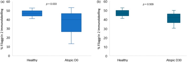
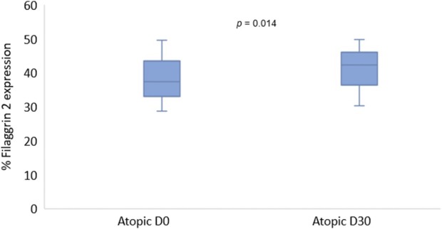
Results: There was a higher FLG2 expression in control skin when compared with atopic skin (lesional and nonlesional) on D0 (p = 0.033). FLG2 expression comparison between control and D30 (nonlesional) did not show a significant difference (p = 0.509). A significant increase in FLG2 expression in atopic nonlesional skin on D30 compared with nonlesional skin on D0 was also observed (p = 0.014).
Conclusions and clinical relevance: Oclacitinib maleate could have a positive impact on cutaneous barrier structure, improving FLG2 expression by decreasing inflammation and cutaneous trauma.
期刊介绍:
Veterinary Dermatology is a bi-monthly, peer-reviewed, international journal which publishes papers on all aspects of the skin of mammals, birds, reptiles, amphibians and fish. Scientific research papers, clinical case reports and reviews covering the following aspects of dermatology will be considered for publication:
-Skin structure (anatomy, histology, ultrastructure)
-Skin function (physiology, biochemistry, pharmacology, immunology, genetics)
-Skin microbiology and parasitology
-Dermatopathology
-Pathogenesis, diagnosis and treatment of skin diseases
-New disease entities


 分享
分享
 求助内容:
求助内容: 应助结果提醒方式:
 扫码关注我们
