Albert K Dadzie, Sabrina P Iddir, Sanjay Ganesh, Behrouz Ebrahimi, Mojtaba Rahimi, Mansour Abtahi, Taeyoon Son, Michael J Heiferman, Xincheng Yao
{"title":"Artificial intelligence in the diagnosis of uveal melanoma: advances and applications.","authors":"Albert K Dadzie, Sabrina P Iddir, Sanjay Ganesh, Behrouz Ebrahimi, Mojtaba Rahimi, Mansour Abtahi, Taeyoon Son, Michael J Heiferman, Xincheng Yao","doi":"10.3389/ebm.2025.10444","DOIUrl":null,"url":null,"abstract":"<p><p>Advancements in machine learning and deep learning have the potential to revolutionize the diagnosis of melanocytic choroidal tumors, including uveal melanoma, a potentially life-threatening eye cancer. Traditional machine learning methods rely heavily on manually selected image features, which can limit diagnostic accuracy and lead to variability in results. In contrast, deep learning models, particularly convolutional neural networks (CNNs), are capable of automatically analyzing medical images, identifying complex patterns, and enhancing diagnostic precision. This review evaluates recent studies that apply machine learning and deep learning approaches to classify uveal melanoma using imaging modalities such as fundus photography, optical coherence tomography (OCT), and ultrasound. The review critically examines each study's research design, methodology, and reported performance metrics, discussing strengths as well as limitations. While fundus photography is the predominant imaging modality being used in current research, integrating multiple imaging techniques, such as OCT and ultrasound, may enhance diagnostic accuracy by combining surface and structural information about the tumor. Key limitations across studies include small dataset sizes, limited external validation, and a reliance on single imaging modalities, all of which restrict model generalizability in clinical settings. Metrics such as accuracy, sensitivity, and area under the curve (AUC) indicate that deep learning models have the potential to outperform traditional methods, supporting their further development for integration into clinical workflows. Future research should aim to address current limitations by developing multimodal models that leverage larger, diverse datasets and rigorous validation, thereby paving the way for more comprehensive, reliable diagnostic tools in ocular oncology.</p>","PeriodicalId":12163,"journal":{"name":"Experimental Biology and Medicine","volume":"250 ","pages":"10444"},"PeriodicalIF":2.7000,"publicationDate":"2025-02-19","publicationTypes":"Journal Article","fieldsOfStudy":null,"isOpenAccess":false,"openAccessPdf":"https://www.ncbi.nlm.nih.gov/pmc/articles/PMC11879745/pdf/","citationCount":"0","resultStr":null,"platform":"Semanticscholar","paperid":null,"PeriodicalName":"Experimental Biology and Medicine","FirstCategoryId":"3","ListUrlMain":"https://doi.org/10.3389/ebm.2025.10444","RegionNum":4,"RegionCategory":"医学","ArticlePicture":[],"TitleCN":null,"AbstractTextCN":null,"PMCID":null,"EPubDate":"2025/1/1 0:00:00","PubModel":"eCollection","JCR":"Q2","JCRName":"MEDICINE, RESEARCH & EXPERIMENTAL","Score":null,"Total":0}
引用次数: 0
Abstract
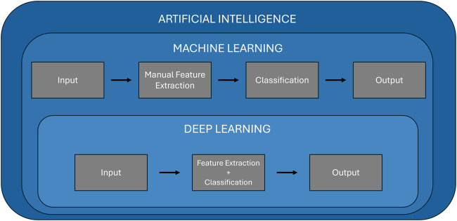
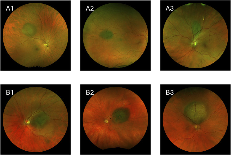
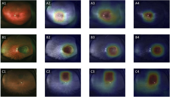
Advancements in machine learning and deep learning have the potential to revolutionize the diagnosis of melanocytic choroidal tumors, including uveal melanoma, a potentially life-threatening eye cancer. Traditional machine learning methods rely heavily on manually selected image features, which can limit diagnostic accuracy and lead to variability in results. In contrast, deep learning models, particularly convolutional neural networks (CNNs), are capable of automatically analyzing medical images, identifying complex patterns, and enhancing diagnostic precision. This review evaluates recent studies that apply machine learning and deep learning approaches to classify uveal melanoma using imaging modalities such as fundus photography, optical coherence tomography (OCT), and ultrasound. The review critically examines each study's research design, methodology, and reported performance metrics, discussing strengths as well as limitations. While fundus photography is the predominant imaging modality being used in current research, integrating multiple imaging techniques, such as OCT and ultrasound, may enhance diagnostic accuracy by combining surface and structural information about the tumor. Key limitations across studies include small dataset sizes, limited external validation, and a reliance on single imaging modalities, all of which restrict model generalizability in clinical settings. Metrics such as accuracy, sensitivity, and area under the curve (AUC) indicate that deep learning models have the potential to outperform traditional methods, supporting their further development for integration into clinical workflows. Future research should aim to address current limitations by developing multimodal models that leverage larger, diverse datasets and rigorous validation, thereby paving the way for more comprehensive, reliable diagnostic tools in ocular oncology.
期刊介绍:
Experimental Biology and Medicine (EBM) is a global, peer-reviewed journal dedicated to the publication of multidisciplinary and interdisciplinary research in the biomedical sciences. EBM provides both research and review articles as well as meeting symposia and brief communications. Articles in EBM represent cutting edge research at the overlapping junctions of the biological, physical and engineering sciences that impact upon the health and welfare of the world''s population.
Topics covered in EBM include: Anatomy/Pathology; Biochemistry and Molecular Biology; Bioimaging; Biomedical Engineering; Bionanoscience; Cell and Developmental Biology; Endocrinology and Nutrition; Environmental Health/Biomarkers/Precision Medicine; Genomics, Proteomics, and Bioinformatics; Immunology/Microbiology/Virology; Mechanisms of Aging; Neuroscience; Pharmacology and Toxicology; Physiology; Stem Cell Biology; Structural Biology; Systems Biology and Microphysiological Systems; and Translational Research.


 分享
分享
 求助内容:
求助内容: 应助结果提醒方式:
 扫码关注我们
