{"title":"Development of a deep learning-based model to evaluate changes during radiotherapy using cervical cancer digital pathology.","authors":"Masaaki Goto, Yasunori Futamura, Hirokazu Makishima, Takashi Saito, Noriaki Sakamoto, Tatsuo Iijima, Yoshio Tamaki, Toshiyuki Okumura, Tetsuya Sakurai, Hideyuki Sakurai","doi":"10.1093/jrr/rraf004","DOIUrl":null,"url":null,"abstract":"<p><p>This study aims to create a deep learning-based classification model for cervical cancer biopsy before and during radiotherapy, visualize the results on whole slide images (WSIs), and explore the clinical significance of obtained features. This study included 95 patients with cervical cancer who received radiotherapy between April 2013 and December 2020. Hematoxylin-eosin stained biopsies were digitized to WSIs and divided into small tiles. Our model adopted the feature extractor of DenseNet121 and the classifier of the support vector machine. About 12 400 tiles were used for training the model and 6000 tiles for testing. The model performance was assessed on a per-tile and per-WSI basis. The resultant probability was defined as radiotherapy status probability (RSP) and its color map was visualized on WSIs. Survival analysis was performed to examine the clinical significance of the RSP. In the test set, the trained model had an area under the receiver operating characteristic curve of 0.76 per-tile and 0.95 per-WSI. In visualization, the model focused on viable tumor components and stroma in tumor biopsies. While survival analysis failed to show the prognostic impact of RSP during treatment, cases with low RSP at diagnosis had prolonged overall survival compared to those with high RSP (P = 0.045). In conclusion, we successfully developed a model to classify biopsies before and during radiotherapy and visualized the result on slide images. Low RSP cases before treatment had a better prognosis, suggesting that tumor morphologic features obtained using the model may be useful for predicting prognosis.</p>","PeriodicalId":16922,"journal":{"name":"Journal of Radiation Research","volume":" ","pages":"144-156"},"PeriodicalIF":2.0000,"publicationDate":"2025-03-24","publicationTypes":"Journal Article","fieldsOfStudy":null,"isOpenAccess":false,"openAccessPdf":"https://www.ncbi.nlm.nih.gov/pmc/articles/PMC11932348/pdf/","citationCount":"0","resultStr":null,"platform":"Semanticscholar","paperid":null,"PeriodicalName":"Journal of Radiation Research","FirstCategoryId":"3","ListUrlMain":"https://doi.org/10.1093/jrr/rraf004","RegionNum":4,"RegionCategory":"医学","ArticlePicture":[],"TitleCN":null,"AbstractTextCN":null,"PMCID":null,"EPubDate":"","PubModel":"","JCR":"Q2","JCRName":"BIOLOGY","Score":null,"Total":0}
引用次数: 0
Abstract
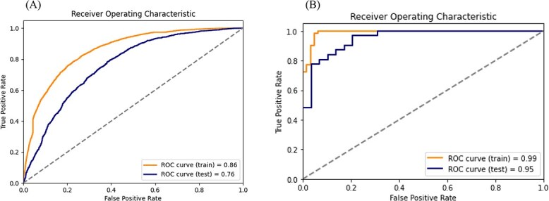
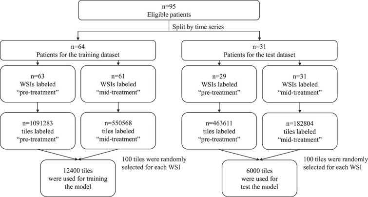
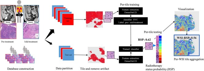
This study aims to create a deep learning-based classification model for cervical cancer biopsy before and during radiotherapy, visualize the results on whole slide images (WSIs), and explore the clinical significance of obtained features. This study included 95 patients with cervical cancer who received radiotherapy between April 2013 and December 2020. Hematoxylin-eosin stained biopsies were digitized to WSIs and divided into small tiles. Our model adopted the feature extractor of DenseNet121 and the classifier of the support vector machine. About 12 400 tiles were used for training the model and 6000 tiles for testing. The model performance was assessed on a per-tile and per-WSI basis. The resultant probability was defined as radiotherapy status probability (RSP) and its color map was visualized on WSIs. Survival analysis was performed to examine the clinical significance of the RSP. In the test set, the trained model had an area under the receiver operating characteristic curve of 0.76 per-tile and 0.95 per-WSI. In visualization, the model focused on viable tumor components and stroma in tumor biopsies. While survival analysis failed to show the prognostic impact of RSP during treatment, cases with low RSP at diagnosis had prolonged overall survival compared to those with high RSP (P = 0.045). In conclusion, we successfully developed a model to classify biopsies before and during radiotherapy and visualized the result on slide images. Low RSP cases before treatment had a better prognosis, suggesting that tumor morphologic features obtained using the model may be useful for predicting prognosis.
期刊介绍:
The Journal of Radiation Research (JRR) is an official journal of The Japanese Radiation Research Society (JRRS), and the Japanese Society for Radiation Oncology (JASTRO).
Since its launch in 1960 as the official journal of the JRRS, the journal has published scientific articles in radiation science in biology, chemistry, physics, epidemiology, and environmental sciences. JRR broadened its scope to include oncology in 2009, when JASTRO partnered with the JRRS to publish the journal.
Articles considered fall into two broad categories:
Oncology & Medicine - including all aspects of research with patients that impacts on the treatment of cancer using radiation. Papers which cover related radiation therapies, radiation dosimetry, and those describing the basis for treatment methods including techniques, are also welcomed. Clinical case reports are not acceptable.
Radiation Research - basic science studies of radiation effects on livings in the area of physics, chemistry, biology, epidemiology and environmental sciences.
Please be advised that JRR does not accept any papers of pure physics or chemistry.
The journal is bimonthly, and is edited and published by the JRR Editorial Committee.


 分享
分享
 求助内容:
求助内容: 应助结果提醒方式:
 扫码关注我们
