{"title":"Kimura disease of the tongue base: a rare case diagnosed through cytological examination of Warthin-Finkeldey-type multinucleated cells.","authors":"Hidetoshi Satomi, Ayumi Ryu, Azusa Shingetsu, Sei Murayama, Yuki Morimoto, Yoshinori Kodama, Satoshi Tanada, Keiichiro Honma","doi":"10.3960/jslrt.25007","DOIUrl":null,"url":null,"abstract":"<p><p>Kimura disease (KD) is a rare chronic inflammatory condition that primarily affects Asian males and typically presents in the head and neck region. We describe an exceptionally rare case of KD involving the lingual tonsil of Waldeyer's ring in a 39-year-old Japanese man, marking only the second reported instance of lingual involvement and the first specifically affecting the tongue base. The patient presented with a well-circumscribed, 3.5-cm mass extending from the lingual tonsil to the epiglottis. Laboratory findings revealed significant peripheral eosinophilia (13.5%) and elevated serum IgE levels (2,750 IU/mL). Because of the challenging location for conventional biopsy, fine-needle aspiration cytology was performed on associated cervical lymph nodes. Cytological examination identified Warthin-Finkeldey-type multinucleated cells, eosinophilic infiltration, and vascular proliferation, leading to a presumptive KD diagnosis based on cytomorphology. The diagnosis was confirmed through surgical excision and histopathological analysis. This case is noteworthy for two reasons: it documents an extremely rare presentation of KD in the tongue base and underscores the diagnostic value of cytological examination in anatomically difficult locations where surgical biopsy may be unfeasible. The presence of Warthin-Finkeldey-type multinucleated cells in cytological specimens provided a key diagnostic clue, particularly when integrated with clinical and laboratory findings. At six months post-surgery, the patient showed no recurrence. This case highlights the importance of considering KD in the differential diagnosis of head and neck masses, even in atypical locations, and demonstrates the potential utility of cytological examination in diagnosing KD.</p>","PeriodicalId":45936,"journal":{"name":"Journal of Clinical and Experimental Hematopathology","volume":" ","pages":"129-134"},"PeriodicalIF":1.4000,"publicationDate":"2025-06-28","publicationTypes":"Journal Article","fieldsOfStudy":null,"isOpenAccess":false,"openAccessPdf":"https://www.ncbi.nlm.nih.gov/pmc/articles/PMC12351233/pdf/","citationCount":"0","resultStr":null,"platform":"Semanticscholar","paperid":null,"PeriodicalName":"Journal of Clinical and Experimental Hematopathology","FirstCategoryId":"1085","ListUrlMain":"https://doi.org/10.3960/jslrt.25007","RegionNum":0,"RegionCategory":null,"ArticlePicture":[],"TitleCN":null,"AbstractTextCN":null,"PMCID":null,"EPubDate":"2025/3/12 0:00:00","PubModel":"Epub","JCR":"Q4","JCRName":"HEMATOLOGY","Score":null,"Total":0}
引用次数: 0
Abstract
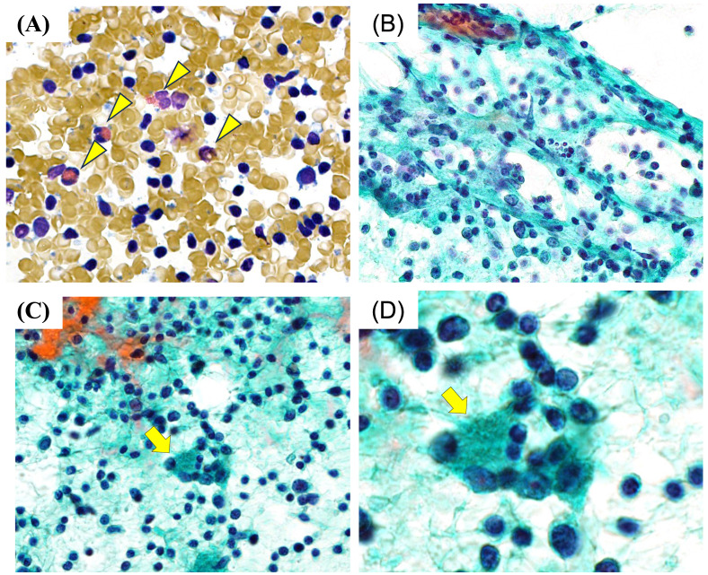
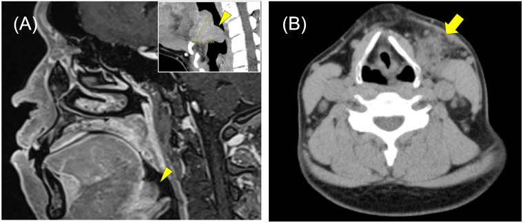
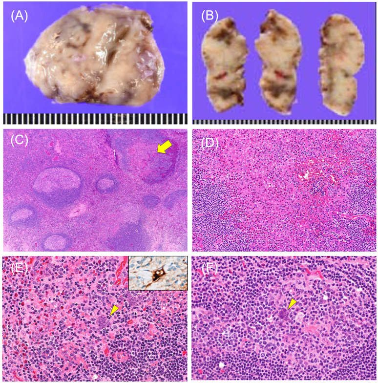
Kimura disease (KD) is a rare chronic inflammatory condition that primarily affects Asian males and typically presents in the head and neck region. We describe an exceptionally rare case of KD involving the lingual tonsil of Waldeyer's ring in a 39-year-old Japanese man, marking only the second reported instance of lingual involvement and the first specifically affecting the tongue base. The patient presented with a well-circumscribed, 3.5-cm mass extending from the lingual tonsil to the epiglottis. Laboratory findings revealed significant peripheral eosinophilia (13.5%) and elevated serum IgE levels (2,750 IU/mL). Because of the challenging location for conventional biopsy, fine-needle aspiration cytology was performed on associated cervical lymph nodes. Cytological examination identified Warthin-Finkeldey-type multinucleated cells, eosinophilic infiltration, and vascular proliferation, leading to a presumptive KD diagnosis based on cytomorphology. The diagnosis was confirmed through surgical excision and histopathological analysis. This case is noteworthy for two reasons: it documents an extremely rare presentation of KD in the tongue base and underscores the diagnostic value of cytological examination in anatomically difficult locations where surgical biopsy may be unfeasible. The presence of Warthin-Finkeldey-type multinucleated cells in cytological specimens provided a key diagnostic clue, particularly when integrated with clinical and laboratory findings. At six months post-surgery, the patient showed no recurrence. This case highlights the importance of considering KD in the differential diagnosis of head and neck masses, even in atypical locations, and demonstrates the potential utility of cytological examination in diagnosing KD.


 分享
分享

 求助内容:
求助内容: 应助结果提醒方式:
 扫码关注我们
