{"title":"Oncocytic Mucoepidermoid Carcinoma of the Parotid Gland.","authors":"Prokopios P Argyris, Paul E Wakely","doi":"10.1007/s12105-025-01768-0","DOIUrl":null,"url":null,"abstract":"<p><strong>Case presentation: </strong>A 30-year-old man presented with a multilobulated left parotid mass measuring 5.2 × 4.1 × 2.4 cm by imaging, and numerous enlarged left cervical lymph nodes, suspicious for metastasis. FNA cytopathology of the mass showed loose clusters of large cells displaying increased N/C ratios and ample granular oncocytic cytoplasm. A superficial left parotidectomy with radical resection of the cheek and cervical lymphadenectomy was performed. Histopathologic examination disclosed a circumscribed, unencapsulated neoplasm exhibiting a solid growth pattern composed of infiltrative islands and nests of cohesive, polygonal, oncocytoid cells in a densely fibrous stroma. Lesional cells exhibited enlarged, oval, open-face nuclei with coarse chromatin and a single acidophilic macronucleolus, voluminous eosinophilic granular cytoplasm and distinct cell membrane borders. Mitotic activity and necrosis were absent. Microcystic architecture was noted solely in a single tumor nest at the periphery. These spaces contained mucinous secretions and were lined by cuboidal oncocytic and intermediate cells with interspersed mucocytes, highlighted by mucicarmine stain. Immunohistochemically, oncocytic cells were strongly and diffusely positive for cytokeratin AE1/AE3, p63 and p40, and uniformly negative for androgen receptor, GATA3, S100, SOX10 and Her-2. A MAML2 rearrangement was identified by FISH, thus confirming the diagnosis of oncocytic variant of mucoepidermoid carcinoma.</p><p><strong>Conclusion: </strong>In this illustrative example, we present the clinicoradiologic, cytologic, histopathologic, and immunophenotypic characteristics of this rare variant of mucoepidermoid carcinoma, together with molecular documentation.</p>","PeriodicalId":47972,"journal":{"name":"Head & Neck Pathology","volume":"19 1","pages":"30"},"PeriodicalIF":4.1000,"publicationDate":"2025-03-15","publicationTypes":"Journal Article","fieldsOfStudy":null,"isOpenAccess":false,"openAccessPdf":"https://www.ncbi.nlm.nih.gov/pmc/articles/PMC11910479/pdf/","citationCount":"0","resultStr":null,"platform":"Semanticscholar","paperid":null,"PeriodicalName":"Head & Neck Pathology","FirstCategoryId":"1085","ListUrlMain":"https://doi.org/10.1007/s12105-025-01768-0","RegionNum":0,"RegionCategory":null,"ArticlePicture":[],"TitleCN":null,"AbstractTextCN":null,"PMCID":null,"EPubDate":"","PubModel":"","JCR":"Q2","JCRName":"PATHOLOGY","Score":null,"Total":0}
引用次数: 0
Abstract
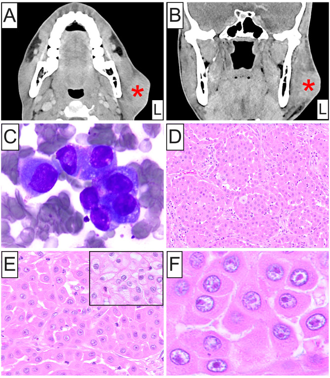
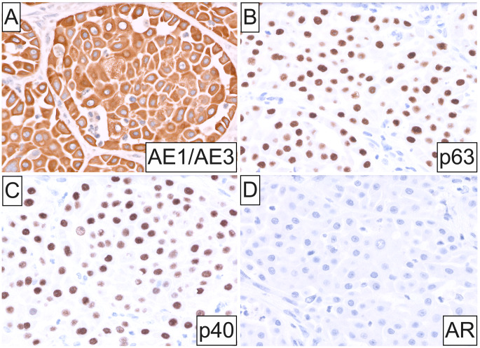
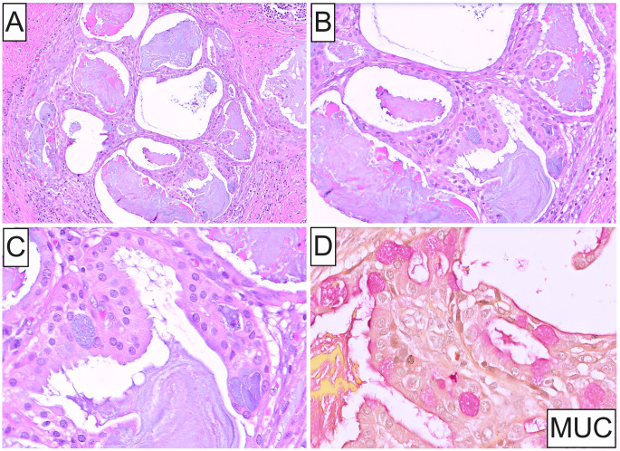
Case presentation: A 30-year-old man presented with a multilobulated left parotid mass measuring 5.2 × 4.1 × 2.4 cm by imaging, and numerous enlarged left cervical lymph nodes, suspicious for metastasis. FNA cytopathology of the mass showed loose clusters of large cells displaying increased N/C ratios and ample granular oncocytic cytoplasm. A superficial left parotidectomy with radical resection of the cheek and cervical lymphadenectomy was performed. Histopathologic examination disclosed a circumscribed, unencapsulated neoplasm exhibiting a solid growth pattern composed of infiltrative islands and nests of cohesive, polygonal, oncocytoid cells in a densely fibrous stroma. Lesional cells exhibited enlarged, oval, open-face nuclei with coarse chromatin and a single acidophilic macronucleolus, voluminous eosinophilic granular cytoplasm and distinct cell membrane borders. Mitotic activity and necrosis were absent. Microcystic architecture was noted solely in a single tumor nest at the periphery. These spaces contained mucinous secretions and were lined by cuboidal oncocytic and intermediate cells with interspersed mucocytes, highlighted by mucicarmine stain. Immunohistochemically, oncocytic cells were strongly and diffusely positive for cytokeratin AE1/AE3, p63 and p40, and uniformly negative for androgen receptor, GATA3, S100, SOX10 and Her-2. A MAML2 rearrangement was identified by FISH, thus confirming the diagnosis of oncocytic variant of mucoepidermoid carcinoma.
Conclusion: In this illustrative example, we present the clinicoradiologic, cytologic, histopathologic, and immunophenotypic characteristics of this rare variant of mucoepidermoid carcinoma, together with molecular documentation.
期刊介绍:
Head & Neck Pathology presents scholarly papers, reviews and symposia that cover the spectrum of human surgical pathology within the anatomic zones of the oral cavity, sinonasal tract, larynx, hypopharynx, salivary gland, ear and temporal bone, and neck.
The journal publishes rapid developments in new diagnostic criteria, intraoperative consultation, immunohistochemical studies, molecular techniques, genetic analyses, diagnostic aids, experimental pathology, cytology, radiographic imaging, and application of uniform terminology to allow practitioners to continue to maintain and expand their knowledge in the subspecialty of head and neck pathology. Coverage of practical application to daily clinical practice is supported with proceedings and symposia from international societies and academies devoted to this field.
Single-blind peer review
The journal follows a single-blind review procedure, where the reviewers are aware of the names and affiliations of the authors, but the reviewer reports provided to authors are anonymous. Single-blind peer review is the traditional model of peer review that many reviewers are comfortable with, and it facilitates a dispassionate critique of a manuscript.


 分享
分享
 求助内容:
求助内容: 应助结果提醒方式:
 扫码关注我们
