Kim Martin, Christos Katsaros, Robert Brylka, Ulrich Schwanecke, Ralf Schulze
{"title":"Influence of patient motion on definition of typical cephalometric reference points in digital horizontally scanning cephalometric radiography.","authors":"Kim Martin, Christos Katsaros, Robert Brylka, Ulrich Schwanecke, Ralf Schulze","doi":"10.1186/s13005-025-00491-z","DOIUrl":null,"url":null,"abstract":"<p><strong>Background: </strong>The aim of this study was to investigate the effect of defined head-motion during x-ray exposure on the identification accuracy of typical cephalometric reference points.</p><p><strong>Methods: </strong>By means of a dry adult human skull and a precise motion simulation system digital Cephs are acquired while certain predefined movement patterns (shift, tilt and nodding with a motion amplitude from 5 - 50 mm) of the skull were executed. They represent the movements of children and adolescents, the main group for cephalometric radiographs.The scanning time was 9.4 s per Ceph. 10 typical landmark points of cephalometric analysis were identified by 20 observers on each Ceph twice. Using a non-motion image (Ceph0) as reference, displacement was computed as vectors relative to this image. Commonly used angles and vertical and horizontal distances were calculated.</p><p><strong>Results: </strong>Both inter-rater as well as intra-rater-reproducibility were perfect. There was very little change in the vertical distance N-Me, in contrast to the horizontal distance S-N which showed a large variation. So patient motion parallel to the scanning direction of the fan-beam-detector unit, heavily influence distances parallel to this direction. The ANB angle and the Maxillo-Mandibular Plane Angle (ANS-PNS to Me-Go) only varied by about 1-2°.</p><p><strong>Conclusions: </strong>The study observed a severe influence on reference point location of motion patterns parallel to the scanning direction and also on clinically relevant distances parallel to the scanning direction. Therefore, we recommend to use a horizontal scanning direction, to minimise scanning time to a minimum, or to prefer a one-shot technique if possible. Future advancements in this field may include the integration of artificial intelligence or algorithms for the purpose of motion correction.</p>","PeriodicalId":12994,"journal":{"name":"Head & Face Medicine","volume":"21 1","pages":"18"},"PeriodicalIF":2.4000,"publicationDate":"2025-03-17","publicationTypes":"Journal Article","fieldsOfStudy":null,"isOpenAccess":false,"openAccessPdf":"https://www.ncbi.nlm.nih.gov/pmc/articles/PMC11912588/pdf/","citationCount":"0","resultStr":null,"platform":"Semanticscholar","paperid":null,"PeriodicalName":"Head & Face Medicine","FirstCategoryId":"3","ListUrlMain":"https://doi.org/10.1186/s13005-025-00491-z","RegionNum":2,"RegionCategory":"医学","ArticlePicture":[],"TitleCN":null,"AbstractTextCN":null,"PMCID":null,"EPubDate":"","PubModel":"","JCR":"Q2","JCRName":"DENTISTRY, ORAL SURGERY & MEDICINE","Score":null,"Total":0}
引用次数: 0
Abstract
Background: The aim of this study was to investigate the effect of defined head-motion during x-ray exposure on the identification accuracy of typical cephalometric reference points.
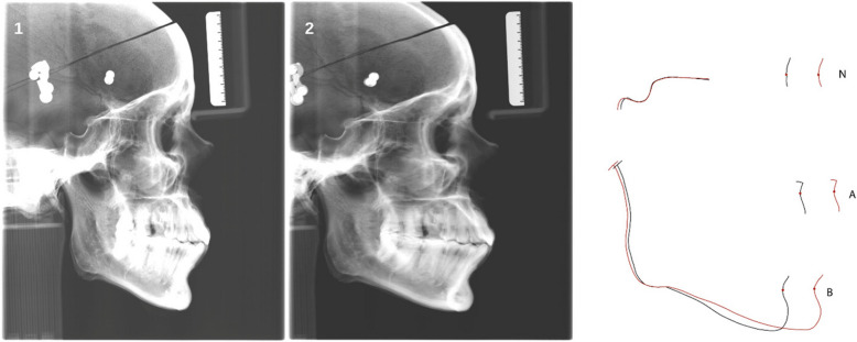
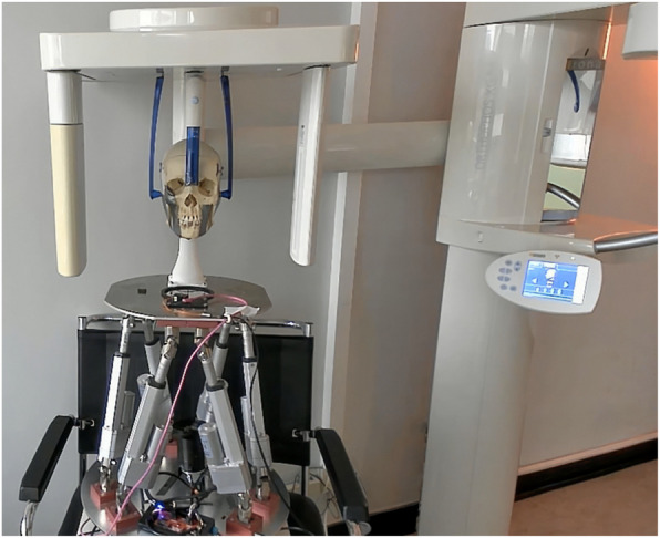
Methods: By means of a dry adult human skull and a precise motion simulation system digital Cephs are acquired while certain predefined movement patterns (shift, tilt and nodding with a motion amplitude from 5 - 50 mm) of the skull were executed. They represent the movements of children and adolescents, the main group for cephalometric radiographs.The scanning time was 9.4 s per Ceph. 10 typical landmark points of cephalometric analysis were identified by 20 observers on each Ceph twice. Using a non-motion image (Ceph0) as reference, displacement was computed as vectors relative to this image. Commonly used angles and vertical and horizontal distances were calculated.
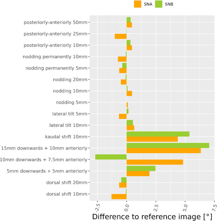
Results: Both inter-rater as well as intra-rater-reproducibility were perfect. There was very little change in the vertical distance N-Me, in contrast to the horizontal distance S-N which showed a large variation. So patient motion parallel to the scanning direction of the fan-beam-detector unit, heavily influence distances parallel to this direction. The ANB angle and the Maxillo-Mandibular Plane Angle (ANS-PNS to Me-Go) only varied by about 1-2°.
Conclusions: The study observed a severe influence on reference point location of motion patterns parallel to the scanning direction and also on clinically relevant distances parallel to the scanning direction. Therefore, we recommend to use a horizontal scanning direction, to minimise scanning time to a minimum, or to prefer a one-shot technique if possible. Future advancements in this field may include the integration of artificial intelligence or algorithms for the purpose of motion correction.
背景:本研究的目的是探讨x线照射时确定的头部运动对典型头颅测量参考点识别准确性的影响,这些参考点是治疗计划的基础。方法:通过干燥的成人颅骨和精确的运动模拟系统,在执行某些预定义的颅骨运动模式(移动,倾斜和点头,运动幅度为5 - 50 mm)时,获得数字Cephs。它们代表儿童和青少年的运动,这是头侧x线片的主要人群。扫描时间为9.4 s / Ceph, 20名观察者在每个Ceph上识别10个典型的头颅测量分析地标点2次。使用非运动图像(Ceph0)作为参考,以相对于该图像的向量计算位移。计算常用角度和垂直、水平距离。结果:组间和组内重现性良好。垂直距离N-Me变化不大,水平距离S-N变化较大。所以病人的运动平行于扇形光束探测器单元的扫描方向,严重影响平行于这个方向的距离。ANB角和上下颌平面角(ANS-PNS to Me-Go)仅变化约1-2°,但足以极大地影响治疗计划。结论:本研究观察到平行于扫描方向的运动模式对参考点位置的严重影响,也对平行于扫描方向的临床相关距离的严重影响。因此,我们建议使用水平扫描方向,以尽量减少扫描时间,或者如果可能的话,首选一次扫描技术。该领域的未来发展可能包括人工智能或运动校正算法的集成。
期刊介绍:
Head & Face Medicine is a multidisciplinary open access journal that publishes basic and clinical research concerning all aspects of cranial, facial and oral conditions.
The journal covers all aspects of cranial, facial and oral diseases and their management. It has been designed as a multidisciplinary journal for clinicians and researchers involved in the diagnostic and therapeutic aspects of diseases which affect the human head and face. The journal is wide-ranging, covering the development, aetiology, epidemiology and therapy of head and face diseases to the basic science that underlies these diseases. Management of head and face diseases includes all aspects of surgical and non-surgical treatments including psychopharmacological therapies.


 分享
分享
 求助内容:
求助内容: 应助结果提醒方式:
 扫码关注我们
