{"title":"Potential of High-Intensity Focused Ultrasound in Enamel Remineralization.","authors":"B Shrestha, S M Rajan, M Saunders, A Fawzy","doi":"10.1177/00220345251323869","DOIUrl":null,"url":null,"abstract":"<p><p>Remineralization is an essential interventional strategy for intercepting enamel white spot lesions (WSLs). Given the limitations of both natural and/or fluoride-mediated repair processes, there is a need to develop novel strategies for repairing enamel WSLs via a minimally invasive approach while restoring the unique ultrastructural integrity and functional properties. Inspired by the unique capability of high-intensity focused ultrasound (HIFU) in facilitating the crystallization process, we propose a novel strategy of employing HIFU for in vitro repair of WSLs through synergizing the crystallization process required for hydroxyapatite (HAP) formation from its precursor (calcium phosphate ion clusters; CPICs). Following CPIC formulation and characterization including the resultant amorphous calcium phosphate (ACP), the effect of HIFU on the ACP-to-HAP transition on the amorphous substrate was investigated using transmission electron microscopy and high-resolution transmission electron microscopy, selected area electron diffraction, and X-ray diffraction (XRD). The results showed profound amorphous-to-crystalline phase transition, within 5- to 30-min HIFU exposure, whereas the long axis of the resultant HAP corresponded with the (002) plane, and a lattice spacing of 0.34 nm indicated a preferred <i>c</i>-axis growth direction consistent with the orientation of natural enamel crystallites. For enamel repair, artificial WSLs were created on enamel specimens and then subjected to CPICs, followed by HIFU exposure for 2.5, 5, or 10 min. Scanning electron and atomic force microscopies revealed the decreased surface roughness and the gradual obliteration in the WSL porous structure with continuous linear coaxial arrangement of HAP crystallites filling the prismatic/interprismatic gaps closely resembling sound enamel specifically with 5-min HIFU exposure. Enamel WSL ultrastructural repair was further confirmed from XRD and Raman spectral analyses with the associated regaining of mineral density and nanomechanical properties as reflected from micro-computed tomography (CT) and nanoindentation results, respectively. Micro-CT further validated the subsurface remineralization of WSLs with HIFU exposure. Within the same exposure parameters, HIFU exhibited a potent antibiofilm effect against <i>Streptococcus mutans</i>. This study introduced a new approach for remineralizing enamel WSLs through the potent synergy between HIFU and CPICs.</p>","PeriodicalId":94075,"journal":{"name":"Journal of dental research","volume":" ","pages":"983-992"},"PeriodicalIF":5.9000,"publicationDate":"2025-08-01","publicationTypes":"Journal Article","fieldsOfStudy":null,"isOpenAccess":false,"openAccessPdf":"https://www.ncbi.nlm.nih.gov/pmc/articles/PMC12209549/pdf/","citationCount":"0","resultStr":null,"platform":"Semanticscholar","paperid":null,"PeriodicalName":"Journal of dental research","FirstCategoryId":"1085","ListUrlMain":"https://doi.org/10.1177/00220345251323869","RegionNum":0,"RegionCategory":null,"ArticlePicture":[],"TitleCN":null,"AbstractTextCN":null,"PMCID":null,"EPubDate":"2025/3/19 0:00:00","PubModel":"Epub","JCR":"","JCRName":"","Score":null,"Total":0}
引用次数: 0
Abstract
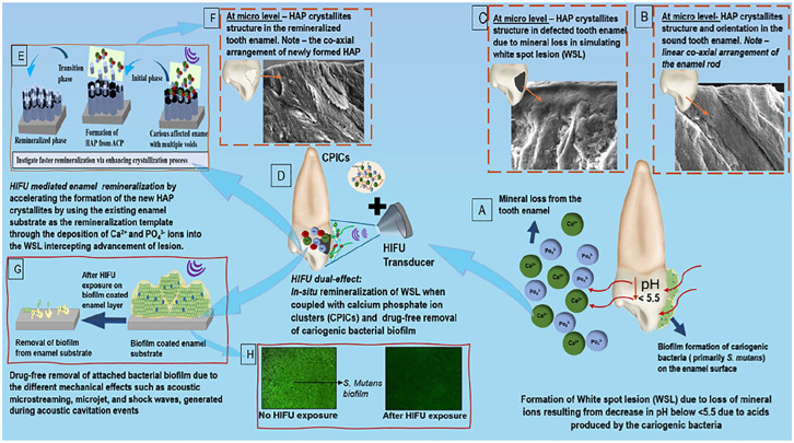
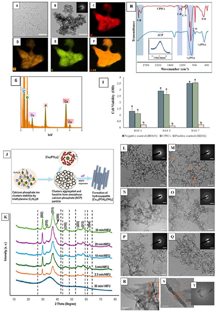
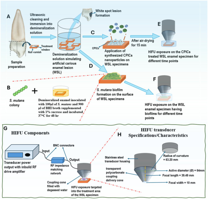
Remineralization is an essential interventional strategy for intercepting enamel white spot lesions (WSLs). Given the limitations of both natural and/or fluoride-mediated repair processes, there is a need to develop novel strategies for repairing enamel WSLs via a minimally invasive approach while restoring the unique ultrastructural integrity and functional properties. Inspired by the unique capability of high-intensity focused ultrasound (HIFU) in facilitating the crystallization process, we propose a novel strategy of employing HIFU for in vitro repair of WSLs through synergizing the crystallization process required for hydroxyapatite (HAP) formation from its precursor (calcium phosphate ion clusters; CPICs). Following CPIC formulation and characterization including the resultant amorphous calcium phosphate (ACP), the effect of HIFU on the ACP-to-HAP transition on the amorphous substrate was investigated using transmission electron microscopy and high-resolution transmission electron microscopy, selected area electron diffraction, and X-ray diffraction (XRD). The results showed profound amorphous-to-crystalline phase transition, within 5- to 30-min HIFU exposure, whereas the long axis of the resultant HAP corresponded with the (002) plane, and a lattice spacing of 0.34 nm indicated a preferred c-axis growth direction consistent with the orientation of natural enamel crystallites. For enamel repair, artificial WSLs were created on enamel specimens and then subjected to CPICs, followed by HIFU exposure for 2.5, 5, or 10 min. Scanning electron and atomic force microscopies revealed the decreased surface roughness and the gradual obliteration in the WSL porous structure with continuous linear coaxial arrangement of HAP crystallites filling the prismatic/interprismatic gaps closely resembling sound enamel specifically with 5-min HIFU exposure. Enamel WSL ultrastructural repair was further confirmed from XRD and Raman spectral analyses with the associated regaining of mineral density and nanomechanical properties as reflected from micro-computed tomography (CT) and nanoindentation results, respectively. Micro-CT further validated the subsurface remineralization of WSLs with HIFU exposure. Within the same exposure parameters, HIFU exhibited a potent antibiofilm effect against Streptococcus mutans. This study introduced a new approach for remineralizing enamel WSLs through the potent synergy between HIFU and CPICs.


 分享
分享

 求助内容:
求助内容: 应助结果提醒方式:
 扫码关注我们
