{"title":"Unexpected vasogenic oedema and alexia as complications after dural arteriovenous fistula embolization.","authors":"Yun-Hsien Ho, Hsin-Fan Chiang, Cheng-Chih Hsieh, Shih-Yang Wei, Chun-Chao Huang","doi":"10.1093/bjrcr/uaaf012","DOIUrl":null,"url":null,"abstract":"<p><p>A 63-year-old male presented with acute onset of intermittent dizziness, visual disturbances, and left temporal headache. Investigations revealed a dural arteriovenous fistula (dAVF) at the left sigmoid sinus, classified as Cognard type IIb. Successful therapeutic transvenous embolization was performed using coils and Onyx, resulting in complete resolution of the dAVF without immediate complications. However, 3 days post-embolization, the patient developed headache, dizziness, visual discomfort, and alexia. MRI findings suggested vasogenic oedema in the left temporo-occipital area due to venous outflow obstruction. Despite treatment with enoxaparin and dexamethasone, the patient experienced progressive symptoms including difficulty in object naming, memory decline, and nonconvulsive seizures. Follow-up imaging indicated improvement of oedema and stable minimal focal gliosis. This rare case of a patient developing alexia following endovascular embolization of a dural AVF highlights the importance of post-procedural monitoring and suggests potential benefits of prophylactic anticoagulation to reduce the risk of probable complications.</p>","PeriodicalId":45216,"journal":{"name":"BJR Case Reports","volume":"11 2","pages":"uaaf012"},"PeriodicalIF":0.5000,"publicationDate":"2025-03-12","publicationTypes":"Journal Article","fieldsOfStudy":null,"isOpenAccess":false,"openAccessPdf":"https://www.ncbi.nlm.nih.gov/pmc/articles/PMC11925498/pdf/","citationCount":"0","resultStr":null,"platform":"Semanticscholar","paperid":null,"PeriodicalName":"BJR Case Reports","FirstCategoryId":"1085","ListUrlMain":"https://doi.org/10.1093/bjrcr/uaaf012","RegionNum":0,"RegionCategory":null,"ArticlePicture":[],"TitleCN":null,"AbstractTextCN":null,"PMCID":null,"EPubDate":"2025/3/1 0:00:00","PubModel":"eCollection","JCR":"Q4","JCRName":"RADIOLOGY, NUCLEAR MEDICINE & MEDICAL IMAGING","Score":null,"Total":0}
引用次数: 0
Abstract
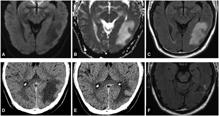
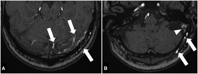
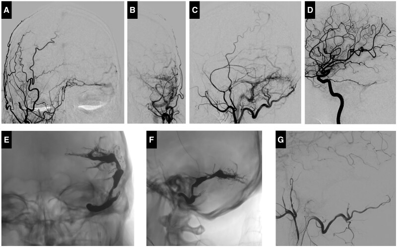
A 63-year-old male presented with acute onset of intermittent dizziness, visual disturbances, and left temporal headache. Investigations revealed a dural arteriovenous fistula (dAVF) at the left sigmoid sinus, classified as Cognard type IIb. Successful therapeutic transvenous embolization was performed using coils and Onyx, resulting in complete resolution of the dAVF without immediate complications. However, 3 days post-embolization, the patient developed headache, dizziness, visual discomfort, and alexia. MRI findings suggested vasogenic oedema in the left temporo-occipital area due to venous outflow obstruction. Despite treatment with enoxaparin and dexamethasone, the patient experienced progressive symptoms including difficulty in object naming, memory decline, and nonconvulsive seizures. Follow-up imaging indicated improvement of oedema and stable minimal focal gliosis. This rare case of a patient developing alexia following endovascular embolization of a dural AVF highlights the importance of post-procedural monitoring and suggests potential benefits of prophylactic anticoagulation to reduce the risk of probable complications.


 分享
分享
 求助内容:
求助内容: 应助结果提醒方式:
 扫码关注我们
