Differentiating Between Fibromuscular Dysplasia and Takayasu Arteritis in a Patient With Juvenile Renovascular Hypertension and Marked Hypokalemia Effectively Treated With Percutaneous Transluminal Renal Angioplasty.
{"title":"Differentiating Between Fibromuscular Dysplasia and Takayasu Arteritis in a Patient With Juvenile Renovascular Hypertension and Marked Hypokalemia Effectively Treated With Percutaneous Transluminal Renal Angioplasty.","authors":"Ayako Ishii, Keisuke Okamura, Shogo Morisaki, Yasunori Momota, Akiko Yamashita, Kenta Hatsuse, Keisuke Kono, Hideto Sako, Akihiro Udo, Kenichiro Taniguchi, Tomoko Koseki, Takuro Arai, Yoshie Yodogawa, Yoshiko Oba, Shiori Hirayama, Miki Inoue, Ichiro Imamura","doi":"10.14740/jocmr6187","DOIUrl":null,"url":null,"abstract":"<p><p>Renovascular hypertension (RVHT) is most commonly caused by renal artery stenosis (RAS) secondary to arteriosclerosis. Other causes of RVHT include fibromuscular dysplasia (FMD) and other rare causes, such as Takayasu arteritis (TA). A male patient in his early 20s presented with hypertension. Laboratory findings were positive for hypokalemia as well as elevations in plasma renin activity and aldosterone concentration. Plain computed tomography revealed atrophy of the right kidney, and magnetic resonance angiography revealed right RAS. A diagnosis of RVHT was suspected, and he was admitted to the cardiovascular ward. After percutaneous transluminal renal angioplasty (PTRA) to treat the right RAS, a typical course was observed with decreased blood pressure, normalizing hypokalemia, and decreased plasma renin activity and aldosterone concentration (which previously were extremely elevated). As angiography showed no remarkable arteriosclerosis of other vessels and given the patient's young age, FMD was suspected as the underlying cause of RVHT. However, the angiographic findings of RAS in the proximal renal artery and the lack of \"string-of-beads\" appearance were atypical for FMD. The patient had chronic inflammation, and further investigation revealed severe stenosis of the right carotid artery. The high C-reactive protein value and the thickened aortic wall in the computed tomography were the suggestive signs for TA. The patient was diagnosed with TA and started on steroid therapy. Although moderate stenosis remained after revascularization of the renal artery in this patient, hypertension improved markedly, demonstrating the effectiveness of PTRA. Given the diagnosis of TA as the underlying disease, the likelihood of recurrent RVHT due to restenosis of the renal artery remains high, and strict follow-up is thus required.</p>","PeriodicalId":94329,"journal":{"name":"Journal of clinical medicine research","volume":"17 3","pages":"174-180"},"PeriodicalIF":2.0000,"publicationDate":"2025-03-01","publicationTypes":"Journal Article","fieldsOfStudy":null,"isOpenAccess":false,"openAccessPdf":"https://www.ncbi.nlm.nih.gov/pmc/articles/PMC11922628/pdf/","citationCount":"0","resultStr":null,"platform":"Semanticscholar","paperid":null,"PeriodicalName":"Journal of clinical medicine research","FirstCategoryId":"1085","ListUrlMain":"https://doi.org/10.14740/jocmr6187","RegionNum":0,"RegionCategory":null,"ArticlePicture":[],"TitleCN":null,"AbstractTextCN":null,"PMCID":null,"EPubDate":"2025/3/9 0:00:00","PubModel":"Epub","JCR":"Q2","JCRName":"MEDICINE, GENERAL & INTERNAL","Score":null,"Total":0}
引用次数: 0
Abstract
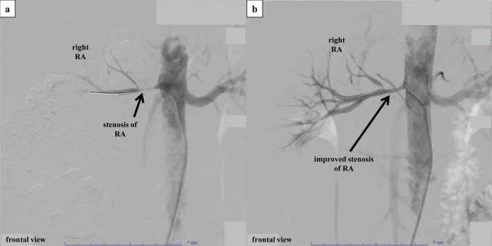
Renovascular hypertension (RVHT) is most commonly caused by renal artery stenosis (RAS) secondary to arteriosclerosis. Other causes of RVHT include fibromuscular dysplasia (FMD) and other rare causes, such as Takayasu arteritis (TA). A male patient in his early 20s presented with hypertension. Laboratory findings were positive for hypokalemia as well as elevations in plasma renin activity and aldosterone concentration. Plain computed tomography revealed atrophy of the right kidney, and magnetic resonance angiography revealed right RAS. A diagnosis of RVHT was suspected, and he was admitted to the cardiovascular ward. After percutaneous transluminal renal angioplasty (PTRA) to treat the right RAS, a typical course was observed with decreased blood pressure, normalizing hypokalemia, and decreased plasma renin activity and aldosterone concentration (which previously were extremely elevated). As angiography showed no remarkable arteriosclerosis of other vessels and given the patient's young age, FMD was suspected as the underlying cause of RVHT. However, the angiographic findings of RAS in the proximal renal artery and the lack of "string-of-beads" appearance were atypical for FMD. The patient had chronic inflammation, and further investigation revealed severe stenosis of the right carotid artery. The high C-reactive protein value and the thickened aortic wall in the computed tomography were the suggestive signs for TA. The patient was diagnosed with TA and started on steroid therapy. Although moderate stenosis remained after revascularization of the renal artery in this patient, hypertension improved markedly, demonstrating the effectiveness of PTRA. Given the diagnosis of TA as the underlying disease, the likelihood of recurrent RVHT due to restenosis of the renal artery remains high, and strict follow-up is thus required.


 分享
分享

 求助内容:
求助内容: 应助结果提醒方式:
 扫码关注我们
