Bong-Hae Cho, Yun-Hoa Jung, Jae-Joon Hwang, Mi-Heon Ryu, Ji-Soo Lee
{"title":"Basal cell adenocarcinoma in the retromolar trigone: A case report.","authors":"Bong-Hae Cho, Yun-Hoa Jung, Jae-Joon Hwang, Mi-Heon Ryu, Ji-Soo Lee","doi":"10.5624/isd.20240226","DOIUrl":null,"url":null,"abstract":"<p><p>Basal cell adenocarcinoma, considered to be the malignant counterpart of basal cell adenoma, is a rare, low-grade malignant tumor of the salivary glands, accounting for 1-2% of salivary gland malignancies. It predominantly affects the parotid gland, while involvement of the minor salivary glands is exceptionally rare. This report presented a case of basal cell adenocarcinoma involving the left retromolar trigone in a 54-year-old woman. The initial provisional diagnosis suggested a benign or low-grade malignant salivary tumor. Advanced magnetic resonance imaging techniques, including diffusion-weighted imaging and apparent diffusion coefficient analysis, aided in the preoperative prediction of malignancy, and an incisional biopsy confirmed the diagnosis of basal cell adenocarcinoma. This case underscored the challenge of distinguishing basal cell adenocarcinoma from benign salivary tumors, as clinical and imaging features often overlap. Surgical excision remains the primary treatment, yielding favorable outcomes; however, long-term follow-up is crucial due to the risk of recurrence.</p>","PeriodicalId":51714,"journal":{"name":"Imaging Science in Dentistry","volume":"55 1","pages":"96-101"},"PeriodicalIF":2.1000,"publicationDate":"2025-03-01","publicationTypes":"Journal Article","fieldsOfStudy":null,"isOpenAccess":false,"openAccessPdf":"https://www.ncbi.nlm.nih.gov/pmc/articles/PMC11966018/pdf/","citationCount":"0","resultStr":null,"platform":"Semanticscholar","paperid":null,"PeriodicalName":"Imaging Science in Dentistry","FirstCategoryId":"1085","ListUrlMain":"https://doi.org/10.5624/isd.20240226","RegionNum":0,"RegionCategory":null,"ArticlePicture":[],"TitleCN":null,"AbstractTextCN":null,"PMCID":null,"EPubDate":"2025/3/10 0:00:00","PubModel":"Epub","JCR":"Q3","JCRName":"DENTISTRY, ORAL SURGERY & MEDICINE","Score":null,"Total":0}
引用次数: 0
Abstract
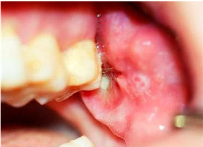
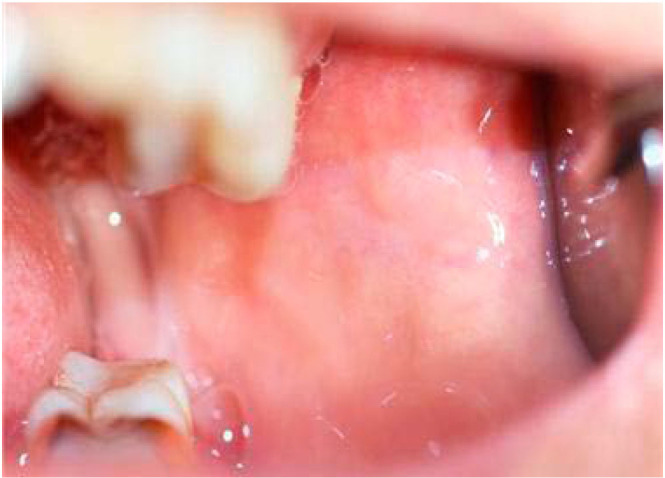
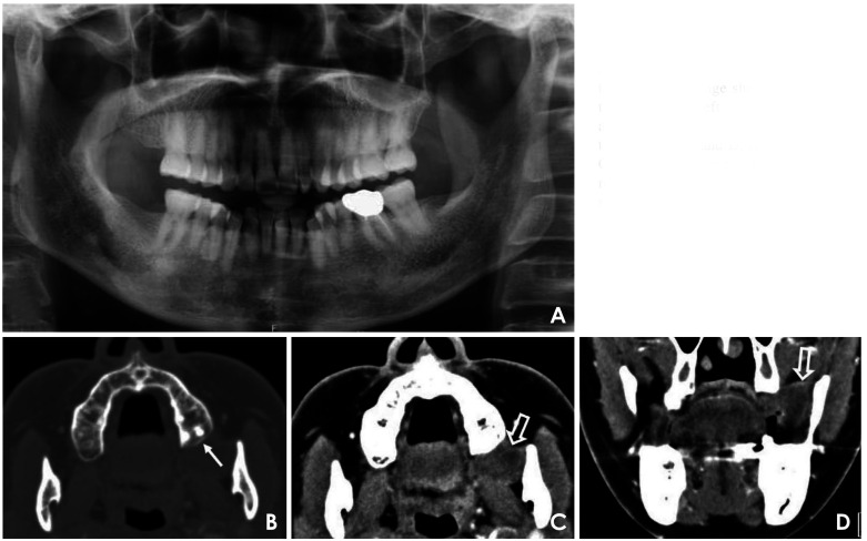
Basal cell adenocarcinoma, considered to be the malignant counterpart of basal cell adenoma, is a rare, low-grade malignant tumor of the salivary glands, accounting for 1-2% of salivary gland malignancies. It predominantly affects the parotid gland, while involvement of the minor salivary glands is exceptionally rare. This report presented a case of basal cell adenocarcinoma involving the left retromolar trigone in a 54-year-old woman. The initial provisional diagnosis suggested a benign or low-grade malignant salivary tumor. Advanced magnetic resonance imaging techniques, including diffusion-weighted imaging and apparent diffusion coefficient analysis, aided in the preoperative prediction of malignancy, and an incisional biopsy confirmed the diagnosis of basal cell adenocarcinoma. This case underscored the challenge of distinguishing basal cell adenocarcinoma from benign salivary tumors, as clinical and imaging features often overlap. Surgical excision remains the primary treatment, yielding favorable outcomes; however, long-term follow-up is crucial due to the risk of recurrence.


 分享
分享
 求助内容:
求助内容: 应助结果提醒方式:
 扫码关注我们
