Jing Wang, Zelong Gu, Longfang Yao, Zhen Zhang, Min Gu
{"title":"Buffer-Free MINFLUX Imaging via Nanobody Points Accumulation","authors":"Jing Wang, Zelong Gu, Longfang Yao, Zhen Zhang, Min Gu","doi":"10.1021/acsphotonics.4c01714","DOIUrl":null,"url":null,"abstract":"MINimal fluorescence photon FLUXes (MINFLUX) nanoscopy, the recently developed super-resolution microscopy method, excels in acquiring high-fidelity images with single-nanometer resolution. The initial implementation of MINFLUX involves separating neighboring fluorophores by individually switching their emission on/off through a complex redox-active blinking buffer, a process found to be operationally cumbersome. While numerous MINFLUX imaging techniques have utilized immobilized probes for nanoscale fluorescence imaging, few have leveraged their binding kinetics. Our study demonstrates the efficacy of MINFLUX imaging facilitated by the accumulation of anti-GFP nanobody at GFP–protein fusions, without the need for additional substances. We explore and adapt nanobody-targeted MINFLUX imaging for diverse subcellular structures. Notably, our approach enables the visualization of the hollow structure of cellular microtubules for the first time in a MINFLUX imaging application. Simultaneously, we performed a comparative analysis of subcellular structure images obtained via STED microscopy. Our labeling technique offers a straightforward and adaptable method for achieving MINFLUX nanoscopy.","PeriodicalId":23,"journal":{"name":"ACS Photonics","volume":"34 1","pages":""},"PeriodicalIF":6.7000,"publicationDate":"2025-04-08","publicationTypes":"Journal Article","fieldsOfStudy":null,"isOpenAccess":false,"openAccessPdf":"","citationCount":"0","resultStr":null,"platform":"Semanticscholar","paperid":null,"PeriodicalName":"ACS Photonics","FirstCategoryId":"101","ListUrlMain":"https://doi.org/10.1021/acsphotonics.4c01714","RegionNum":1,"RegionCategory":"物理与天体物理","ArticlePicture":[],"TitleCN":null,"AbstractTextCN":null,"PMCID":null,"EPubDate":"","PubModel":"","JCR":"Q1","JCRName":"MATERIALS SCIENCE, MULTIDISCIPLINARY","Score":null,"Total":0}
引用次数: 0
Abstract
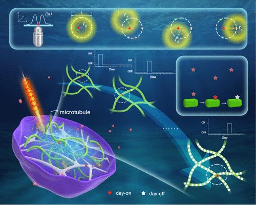
MINimal fluorescence photon FLUXes (MINFLUX) nanoscopy, the recently developed super-resolution microscopy method, excels in acquiring high-fidelity images with single-nanometer resolution. The initial implementation of MINFLUX involves separating neighboring fluorophores by individually switching their emission on/off through a complex redox-active blinking buffer, a process found to be operationally cumbersome. While numerous MINFLUX imaging techniques have utilized immobilized probes for nanoscale fluorescence imaging, few have leveraged their binding kinetics. Our study demonstrates the efficacy of MINFLUX imaging facilitated by the accumulation of anti-GFP nanobody at GFP–protein fusions, without the need for additional substances. We explore and adapt nanobody-targeted MINFLUX imaging for diverse subcellular structures. Notably, our approach enables the visualization of the hollow structure of cellular microtubules for the first time in a MINFLUX imaging application. Simultaneously, we performed a comparative analysis of subcellular structure images obtained via STED microscopy. Our labeling technique offers a straightforward and adaptable method for achieving MINFLUX nanoscopy.
期刊介绍:
Published as soon as accepted and summarized in monthly issues, ACS Photonics will publish Research Articles, Letters, Perspectives, and Reviews, to encompass the full scope of published research in this field.


 分享
分享
 求助内容:
求助内容: 应助结果提醒方式:
 扫码关注我们
