Claire A. Dessalles, Nicolas Cuny, Arthur Boutillon, Paul F. Salipante, Avin Babataheri, Abdul I. Barakat, Guillaume Salbreux
{"title":"Interplay of actin nematodynamics and anisotropic tension controls endothelial mechanics","authors":"Claire A. Dessalles, Nicolas Cuny, Arthur Boutillon, Paul F. Salipante, Avin Babataheri, Abdul I. Barakat, Guillaume Salbreux","doi":"10.1038/s41567-025-02847-3","DOIUrl":null,"url":null,"abstract":"Blood vessels expand and contract actively as they continuously experience dynamic external stresses from blood flow. The mechanical response of the vessel wall is that of a composite material: its mechanical properties depend on its cellular components, which change dynamically as the cells respond to external stress. Mapping the relationship between these underlying cellular processes and emergent tissue mechanics is an ongoing challenge, particularly in endothelial cells. Here we assess the mechanics and cellular dynamics of an endothelial tube using a microstretcher that mimics the native environment of blood vessels. The characterization of the instantaneous monolayer elasticity reveals a strain-stiffening, actin-dependent and substrate-responsive behaviour. After a physiological pressure increase, the tissue displays a fluid-like expansion, with the reorientation of cell shape and actin fibres. We introduce a mechanical model that considers the actin fibres as a network in the nematic phase and couples their dynamics with active and elastic fibre tension. The model accurately describes the response to the pressure of endothelial tubes. Blood flow through a vessel deforms vessel walls. Cells lining these walls sense the changes in pressure as blood flows and reorient their actin fibres in the direction of largest tension.","PeriodicalId":19100,"journal":{"name":"Nature Physics","volume":"21 6","pages":"999-1008"},"PeriodicalIF":18.4000,"publicationDate":"2025-04-18","publicationTypes":"Journal Article","fieldsOfStudy":null,"isOpenAccess":false,"openAccessPdf":"https://www.nature.comhttps://www.nature.com/articles/s41567-025-02847-3.pdf","citationCount":"0","resultStr":null,"platform":"Semanticscholar","paperid":null,"PeriodicalName":"Nature Physics","FirstCategoryId":"101","ListUrlMain":"https://www.nature.com/articles/s41567-025-02847-3","RegionNum":1,"RegionCategory":"物理与天体物理","ArticlePicture":[],"TitleCN":null,"AbstractTextCN":null,"PMCID":null,"EPubDate":"","PubModel":"","JCR":"Q1","JCRName":"PHYSICS, MULTIDISCIPLINARY","Score":null,"Total":0}
引用次数: 0
Abstract
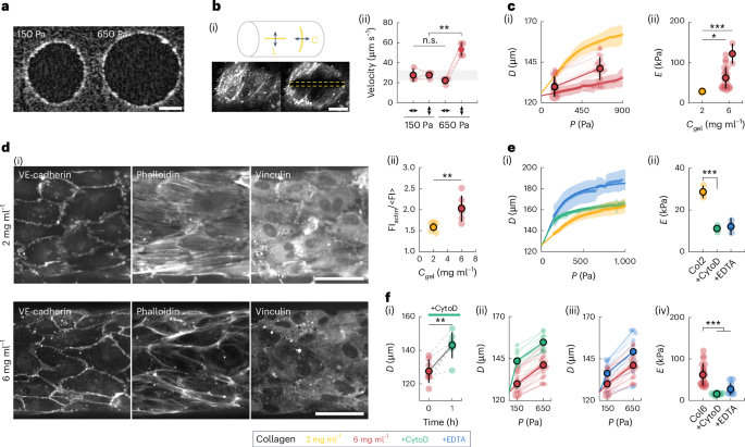
Blood vessels expand and contract actively as they continuously experience dynamic external stresses from blood flow. The mechanical response of the vessel wall is that of a composite material: its mechanical properties depend on its cellular components, which change dynamically as the cells respond to external stress. Mapping the relationship between these underlying cellular processes and emergent tissue mechanics is an ongoing challenge, particularly in endothelial cells. Here we assess the mechanics and cellular dynamics of an endothelial tube using a microstretcher that mimics the native environment of blood vessels. The characterization of the instantaneous monolayer elasticity reveals a strain-stiffening, actin-dependent and substrate-responsive behaviour. After a physiological pressure increase, the tissue displays a fluid-like expansion, with the reorientation of cell shape and actin fibres. We introduce a mechanical model that considers the actin fibres as a network in the nematic phase and couples their dynamics with active and elastic fibre tension. The model accurately describes the response to the pressure of endothelial tubes. Blood flow through a vessel deforms vessel walls. Cells lining these walls sense the changes in pressure as blood flows and reorient their actin fibres in the direction of largest tension.
期刊介绍:
Nature Physics is dedicated to publishing top-tier original research in physics with a fair and rigorous review process. It provides high visibility and access to a broad readership, maintaining high standards in copy editing and production, ensuring rapid publication, and maintaining independence from academic societies and other vested interests.
The journal presents two main research paper formats: Letters and Articles. Alongside primary research, Nature Physics serves as a central source for valuable information within the physics community through Review Articles, News & Views, Research Highlights covering crucial developments across the physics literature, Commentaries, Book Reviews, and Correspondence.


 分享
分享
 求助内容:
求助内容: 应助结果提醒方式:
 扫码关注我们
