{"title":"Developmental fates of shark head cavities reveal mesodermal contributions to tendon progenitor cells in extraocular muscles.","authors":"Shunya Kuroda, Noritaka Adachi, Rie Kusakabe, Shigeru Kuratani","doi":"10.1186/s40851-021-00170-2","DOIUrl":null,"url":null,"abstract":"<p><p>Vertebrate extraocular muscles (EOMs) function in eye movements. The EOMs of modern jawed vertebrates consist primarily of four recti and two oblique muscles innervated by three cranial nerves. The developmental mechanisms underlying the establishment of this complex and the evolutionarily conserved pattern of EOMs are unknown. Chondrichthyan early embryos develop three pairs of overt epithelial coeloms called head cavities (HCs) in the head mesoderm, and each HC is believed to differentiate into a discrete subset of EOMs. However, no direct evidence of these cell fates has been provided due to the technical difficulty of lineage tracing experiments in chondrichthyans. Here, we set up an in ovo manipulation system for embryos of the cloudy catshark Scyliorhinus torazame and labeled the epithelial cells of each HC with lipophilic fluorescent dyes. This experimental system allowed us to trace the cell lineage of EOMs with the highest degree of detail and reproducibility to date. We confirmed that the HCs are indeed primordia of EOMs but showed that the morphological pattern of shark EOMs is not solely dependent on the early pattern of the head mesoderm, which transiently appears as tripartite HCs along the simple anteroposterior axis. Moreover, we found that one of the HCs gives rise to tendon progenitor cells of the EOMs, which is an exceptional condition in our previous understanding of head muscles; the tendons associated with head muscles have generally been supposed to be derived from cranial neural crest (CNC) cells, another source of vertebrate head mesenchyme. Based on interspecies comparisons, the developmental environment is suggested to be significantly different between the two ends of the rectus muscles, and this difference is suggested to be evolutionarily conserved in jawed vertebrates. We propose that the mesenchymal interface (head mesoderm vs CNC) in the environment of developing EOM is required to determine the processes of the proximodistal axis of rectus components of EOMs.</p>","PeriodicalId":54280,"journal":{"name":"Zoological Letters","volume":"7 1","pages":"3"},"PeriodicalIF":1.7000,"publicationDate":"2021-02-15","publicationTypes":"Journal Article","fieldsOfStudy":null,"isOpenAccess":false,"openAccessPdf":"https://sci-hub-pdf.com/10.1186/s40851-021-00170-2","citationCount":"5","resultStr":null,"platform":"Semanticscholar","paperid":null,"PeriodicalName":"Zoological Letters","FirstCategoryId":"99","ListUrlMain":"https://doi.org/10.1186/s40851-021-00170-2","RegionNum":3,"RegionCategory":"生物学","ArticlePicture":[],"TitleCN":null,"AbstractTextCN":null,"PMCID":null,"EPubDate":"","PubModel":"","JCR":"Q2","JCRName":"ZOOLOGY","Score":null,"Total":0}
引用次数: 5
Abstract
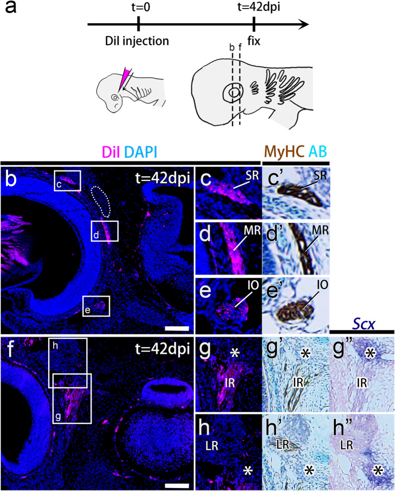
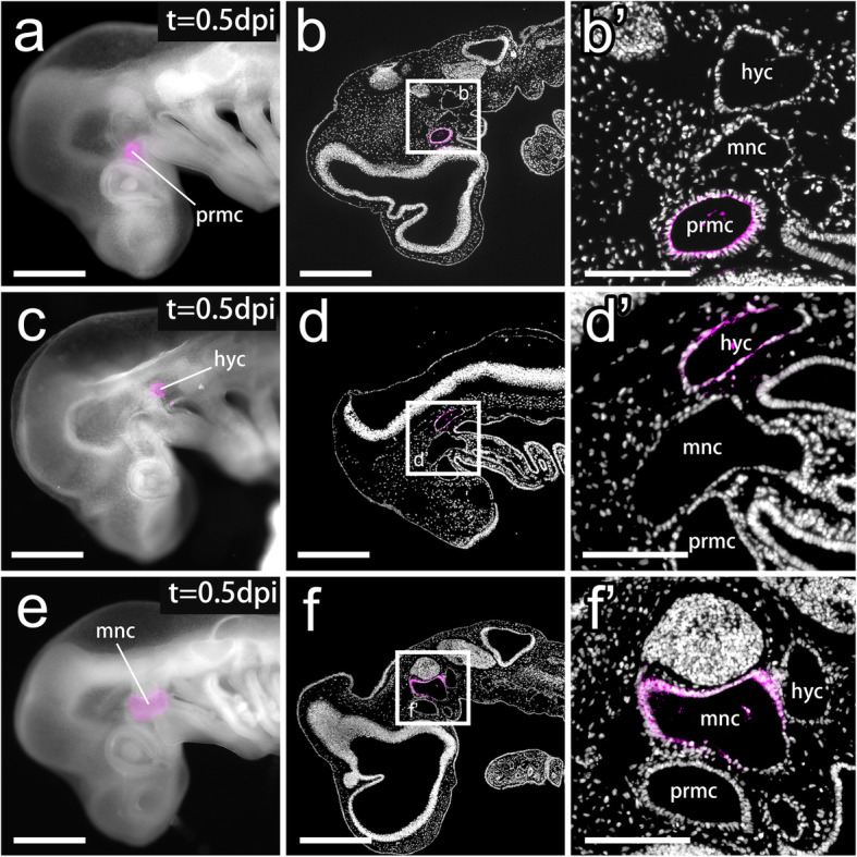
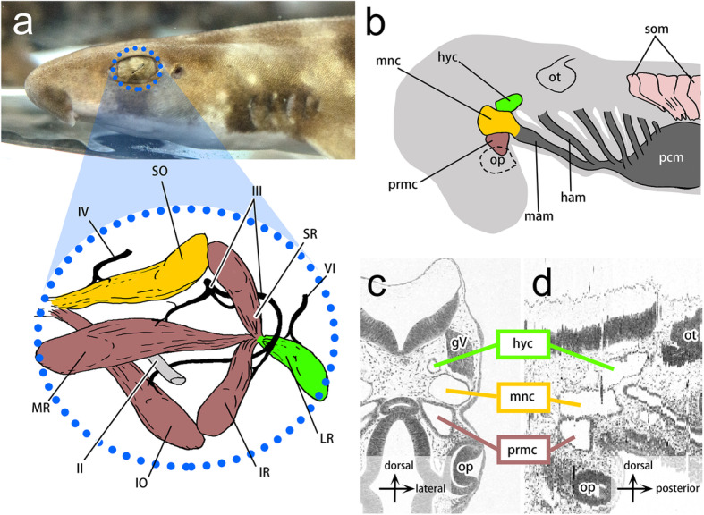
Vertebrate extraocular muscles (EOMs) function in eye movements. The EOMs of modern jawed vertebrates consist primarily of four recti and two oblique muscles innervated by three cranial nerves. The developmental mechanisms underlying the establishment of this complex and the evolutionarily conserved pattern of EOMs are unknown. Chondrichthyan early embryos develop three pairs of overt epithelial coeloms called head cavities (HCs) in the head mesoderm, and each HC is believed to differentiate into a discrete subset of EOMs. However, no direct evidence of these cell fates has been provided due to the technical difficulty of lineage tracing experiments in chondrichthyans. Here, we set up an in ovo manipulation system for embryos of the cloudy catshark Scyliorhinus torazame and labeled the epithelial cells of each HC with lipophilic fluorescent dyes. This experimental system allowed us to trace the cell lineage of EOMs with the highest degree of detail and reproducibility to date. We confirmed that the HCs are indeed primordia of EOMs but showed that the morphological pattern of shark EOMs is not solely dependent on the early pattern of the head mesoderm, which transiently appears as tripartite HCs along the simple anteroposterior axis. Moreover, we found that one of the HCs gives rise to tendon progenitor cells of the EOMs, which is an exceptional condition in our previous understanding of head muscles; the tendons associated with head muscles have generally been supposed to be derived from cranial neural crest (CNC) cells, another source of vertebrate head mesenchyme. Based on interspecies comparisons, the developmental environment is suggested to be significantly different between the two ends of the rectus muscles, and this difference is suggested to be evolutionarily conserved in jawed vertebrates. We propose that the mesenchymal interface (head mesoderm vs CNC) in the environment of developing EOM is required to determine the processes of the proximodistal axis of rectus components of EOMs.
Zoological LettersAgricultural and Biological Sciences-Animal Science and Zoology
CiteScore
3.60
自引率
0.00%
发文量
12
审稿时长
10 weeks
期刊介绍:
Zoological Letters is an open access journal that publishes new and important findings in the zoological sciences. As a sister journal to Zoological Science, Zoological Letters covers a wide range of basic fields of zoology, from taxonomy to bioinformatics. We also welcome submissions of paleontology reports as part of our effort to contribute to the development of new perspectives in evolutionary zoology. Our goal is to serve as a global publishing forum for fundamental researchers in all fields of zoology.


 分享
分享
 求助内容:
求助内容: 应助结果提醒方式:
 扫码关注我们
