{"title":"Radiographic Evaluation of Infraorbital Canal Protrusion into Maxillary Sinus Using Cone-Beam Computed Tomography.","authors":"Fahrettin Kalabalık, Tunahan Aktaş, Ender Akan, Emre Aytuğar","doi":"10.5037/jomr.2020.11405","DOIUrl":null,"url":null,"abstract":"<p><strong>Objectives: </strong>The aim of this retrospective study was to evaluate the relation of the infraorbital canal course with the maxillary sinus using cone-beam computed tomography.</p><p><strong>Material and methods: </strong>A total of 1000 infraorbital canals (IOC) were examined from 500 cone-beam computed tomography scans. IOCs were classified into three types based on the degree of protrusion into the sinus. The presence of Haller cells and mucosal thickening in the sinus were evaluated. The length of bony septum from the canal to the sinus wall (D1), the distance at which protrusion begins posterior to the inferior orbital rim (D2), the vertical distance from the canal to the sinus roof (D3), and the vertical distance from the canal to the sinus floor (D4) were measured.</p><p><strong>Results: </strong>The prevalence of IOC protrusion into the sinus was 8.8%. There was a significant difference in the prevalence of Haller cells between IOC types (P < 0.01). However, no significant correlation was found between IOC types and the presence of mucosal thickening (P > 0.05). There was no significant difference in the mean D1, D2, and D3 between the genders (P > 0.05). The mean D4 was significantly higher in males than in females (P < 0.05).</p><p><strong>Conclusions: </strong>The protrusion of infraorbital canals into the sinus is a common variation that must be considered to prevent accidental injury. Our findings suggest that the risk of injury to the descending canals is very low during routine dentoalveolar procedures because the protruded canal is not close to the sinus floor.</p>","PeriodicalId":230885,"journal":{"name":"Journal of Oral & Maxillofacial Research","volume":" ","pages":"e5"},"PeriodicalIF":0.0000,"publicationDate":"2020-12-31","publicationTypes":"Journal Article","fieldsOfStudy":null,"isOpenAccess":false,"openAccessPdf":"https://ftp.ncbi.nlm.nih.gov/pub/pmc/oa_pdf/17/1d/jomr-11-e5.PMC7875100.pdf","citationCount":"1","resultStr":null,"platform":"Semanticscholar","paperid":null,"PeriodicalName":"Journal of Oral & Maxillofacial Research","FirstCategoryId":"1085","ListUrlMain":"https://doi.org/10.5037/jomr.2020.11405","RegionNum":0,"RegionCategory":null,"ArticlePicture":[],"TitleCN":null,"AbstractTextCN":null,"PMCID":null,"EPubDate":"2020/10/1 0:00:00","PubModel":"eCollection","JCR":"","JCRName":"","Score":null,"Total":0}
引用次数: 1
Abstract
Objectives: The aim of this retrospective study was to evaluate the relation of the infraorbital canal course with the maxillary sinus using cone-beam computed tomography.
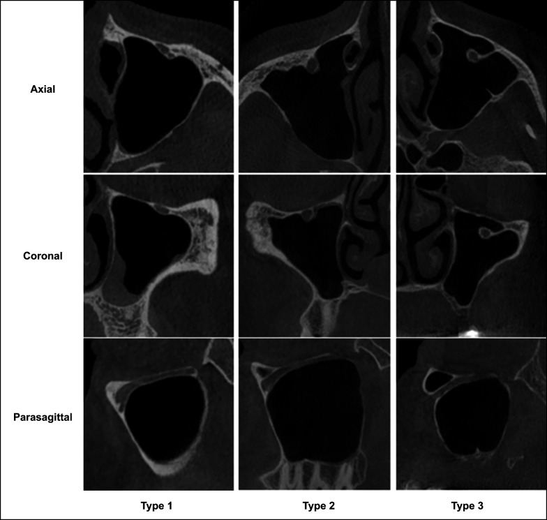
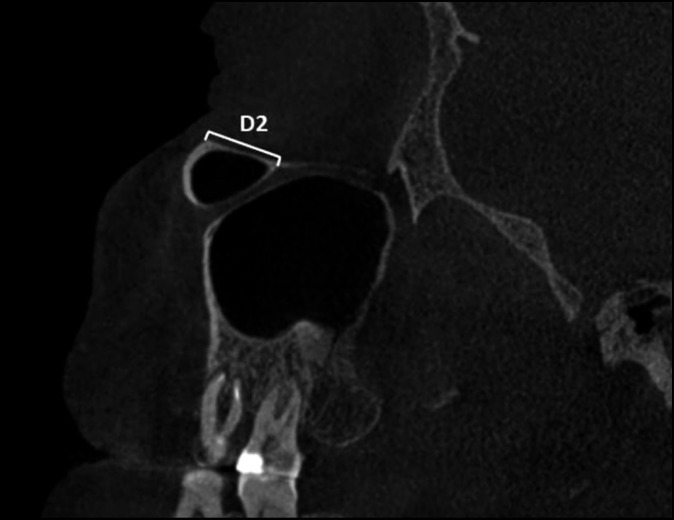
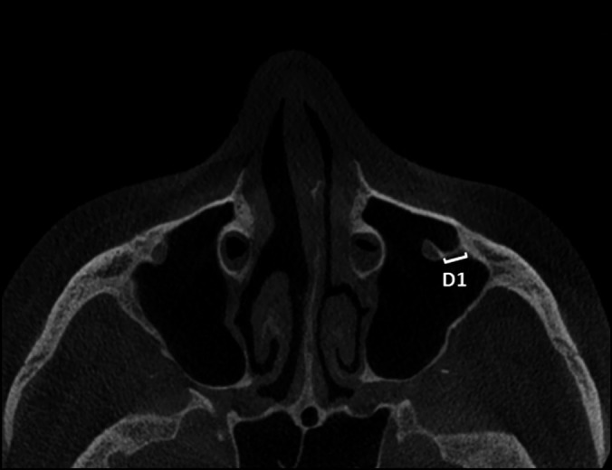
Material and methods: A total of 1000 infraorbital canals (IOC) were examined from 500 cone-beam computed tomography scans. IOCs were classified into three types based on the degree of protrusion into the sinus. The presence of Haller cells and mucosal thickening in the sinus were evaluated. The length of bony septum from the canal to the sinus wall (D1), the distance at which protrusion begins posterior to the inferior orbital rim (D2), the vertical distance from the canal to the sinus roof (D3), and the vertical distance from the canal to the sinus floor (D4) were measured.
Results: The prevalence of IOC protrusion into the sinus was 8.8%. There was a significant difference in the prevalence of Haller cells between IOC types (P < 0.01). However, no significant correlation was found between IOC types and the presence of mucosal thickening (P > 0.05). There was no significant difference in the mean D1, D2, and D3 between the genders (P > 0.05). The mean D4 was significantly higher in males than in females (P < 0.05).
Conclusions: The protrusion of infraorbital canals into the sinus is a common variation that must be considered to prevent accidental injury. Our findings suggest that the risk of injury to the descending canals is very low during routine dentoalveolar procedures because the protruded canal is not close to the sinus floor.


 分享
分享

 求助内容:
求助内容: 应助结果提醒方式:
 扫码关注我们
