{"title":"Microfluidic Applications for Andrology","authors":"Ronald Suh, Shuichi Takayama, Gary D. Smith","doi":"10.2164/jandrol.05119","DOIUrl":null,"url":null,"abstract":"<p>Perhaps one of the most exciting and revolutionary scientific discoveries of the past 3 decades has been the development of in vitro fertilization (IVF) to treat human infertility. It is impossible to quantify its effect on numerous families since the first IVF birth in 1978 in Old-ham, England (Steptoe and Edwards, 1978). With increasing clinical utilization of assisted reproductive technologies (ART), scientists and clinicians gain insights into basic gamete and embryo biology and translate that knowledge into improving the process of IVF. Critical analyses of individual steps have improved outcomes. Attention to sperm processing and isolation to increase recovery of motile sperm and reduce sperm damage has improved fertilization rates and embryo development (Mortimer, 1994). Use of intracytoplasmic sperm injection (ICSI) has allowed fertilization even in severe cases of compromised sperm quality or number (Bonduelle et al, 1999). Finally, refinement of embryo culture has led to improved in vitro embryo development and implantation rates (Gardner and Lane, 1998; Pool, 2002). Most scientific attention, however, has focused on methodologies rather than technology development and equipment. Semen is still processed in test tubes regardless of technique, sperm are physically placed with oocytes after processing, and fertilization and embryo culture occur in culture dishes, test tubes, or both with relatively large volumes (Trounson and Gardner, 2000). With the exception of gamete and embryo micromanipulation, no technologic advancements in IVF have reached widespread use. Nevertheless, it is precisely those technologic advancements, rather than procedural or methodology changes, that have had the greatest effect on assisted reproduction.</p><p>A promising new technology, microfluidics, exists and is becoming increasingly studied. This technology shows promise as an alternative for each step in the IVF process. Microfluidics, based on physical principles of fluid behavior in a microenvironment, has been used widely in chemistry and molecular biology applications (Tomlinson et al, 1995). Currently, microfluidics is gaining interest in studies of cellular behavior and interactions (Shim et al, 2003). In this article, we introduce basics of fluid behavior at the microscale and highlight previous uses of this technology outside of the reproductive sciences. We then describe fabrication of devices and review initial studies that used microfluidics in sperm sorting and microinsemination. Last, we point out some limitations of this new technology and provide speculation on future directions and application of microfluidics in ART.</p><p>Fluid mechanics is a complex physical and mathematical science; therefore, an extensive technical description and review of fluid physics is beyond the scope and intent of this review. Instead, basic principles will be discussed that govern fluid behavior in a microenvironment, especially those aspects with a specific link to devices and technology currently being developed for IVF. We have purposely avoided including mathematical details, choosing instead to convey a general conceptual sense of fluid mechanics present within microchannels. A comprehensive technical and mathematical description of microfluidic physics can be found in excellent reviews from Beebe et al (2002a) and Brody et al (1996).</p><p>Fluids at the microscale are subject to forces typically not important at scales present in our everyday lives. Fluid at the scale of our normal environment is turbulent; particles within a stream of fluid move in an unpredictable pattern. Turbulent flow depends on certain fluid characteristics (viscosity, density, and velocity) and the geometry and size of the channel, leading to calculation of a value known as the Reynold's number. As the scale of the channel reaches micrometer levels, the Reynold's number decreases and becomes increasingly dependent on fluid characteristics. Decrease of the Reynold's number below a threshold value leads to fluid flow in a laminar fashion. Simply put, flow within microchannels becomes streamlined and predictable (Figure 1). At the microscale, fluid behavior becomes increasingly governed by viscous forces and surface tension, which can be described as the cohesiveness of the liquid's molecules.</p><p>This dominance by viscous forces results in several interesting phenomena. Flows with a low Reynold's number possess little to no momentum; thus, fluids within a microchannel respond quickly and reliably to changes in external forces. In addition, at the microscale, 2 or more streams of laminar flow in contact with each other do not mix, except by diffusion of molecules across the interface of the streams. The rate of diffusion between the contacting surfaces at the microscale can be very quick, partially because of the relatively short distances needed to cross fluid volumes.</p><p>Many of these fluid characteristics at the microscale form the principles driving the interest in the use of microchannels for gamete and embryo manipulation. In general, a microenvironment more closely resembles the in vivo conditions of fertilization and development when compared with a culture dish or drop of media. Below, we discuss the theory behind investigating the use of microfluidics in andrology, its testing, limitation, and potential future influence.</p><p>Interest in microfluidics began with attempts to miniaturize chemical and biological analysis devices in the laboratory (Kricka, 1998). Current designs are often referred to as “laboratory-on-a-chip” or micrototal analysis systems (μTAS) and function by allowing a variety of chemical processes and interactions to occur as fluid flows within their miniature channels and chambers (Weigl and Yager, 1999). Such devices perform all the analytical functions necessary for their purpose, including sample handling, mixing, incubation, sorting, transport, interaction, and detection or signaling within an integrated microfluidic “chip.” Examples include, but are not limited to, immunoassays for antibodies present in serum (Linder et al, 2002) and assays determining enzyme reaction kinetics (Xue et al, 2001; Yakovleva et al, 2002).</p><p>Additional applications in cellular biology have emerged, such as integrated cell sorting devices working at the microscale (Fu et al, 2002) and microfluidic devices that allow for the study of cellular interactions with substrates or other cells (Shim et al, 2003). Advances in cell biology have been demonstrated with the use of microfluidics and the principle of laminar flow, allowing for selective exposure of subcellular areas of interest to membrane-permeable molecules (Takayama et al, 2001). Such precise delivery of molecules to cellular subdomains illustrates the precision with which microfluidic regulation of fluid flow is capable.</p><p>Advantages of such laboratory-on-a-chip technology are multiple. First, once designed and tested, the manufacture of such devices is straightforward and inexpensive, allowing them to be disposable (McDonald et al, 2000). Microfluidic analysis devices use very low volumes of samples and reagents and provide for faster reactions and response times (Weigl and Yager, 1999). Miniaturization very importantly allows for integration of multiple processes within a small, self-contained unit (Kricka, 1998). This can be translated into either multiple parallel analyses, consecutive serial processes, or both.</p><p>The brief overview given here is only intended to familiarize readers with the variety of capabilities of microfluidic technology and is by no means a comprehensive listing of microfluidic applications in sciences. Readers are encouraged to consult more thorough reviews (Khandurina and Guttman, 2002; Verpoorte, 2002).</p><p>Microfluidics systems were initially fabricated with the use of materials and techniques common in the industry that inspired them—microelectronics (McDonald et al, 2000). Photolithography and etching of silicon and glass was a highly developed technology also readily available to researchers interested in miniaturizing analytical systems, yet costs were a significant barrier. In search of a suitable alternative, polymers have quickly emerged as a material for microfluidic biological device fabrication (McDonald et al, 2000). Compounds such as poly(methyl)methacrylate (Martynova et al, 1997), fluorinated ethylene propylene (Sahlin et al, 2002), and poly(dimethylsiloxane) (PDMS; McDonald and Whitesides, 2002) are cheaper and easier to manipulate than silicon-glass alternatives (Martynova et al, 1997). PDMS in particular has become one of the most actively explored and promising materials thus far, possessing numerous characteristics specifically suitable for biological use. It is nontoxic, transparent, insulating, and permeable to gases (McDonald and Whitesides, 2002). From a fabrication standpoint, PDMS permits submicron fidelity with molding, cures at low temperatures, and can easily seal reversibly to itself and a host of other materials (McDonald et al, 2000).</p><p>Although PDMS is generally regarded as nontoxic, special consideration must be given to its use with gametes and embryos, which can be very sensitive to their environment compared with transformed cell lines. Before the use of microfluidic devices with sperm, testing confirmed that no negative effects resulted from prolonged exposure to the materials used in their fabrication. Schuster et al (2003) reported that 30 minutes of exposure to PDMS did not alter sperm survival. In addition, Glasgow et al (2001) found that development of 2-cell mouse embryos to the blastocyst stage was unchanged by continuous exposure to numerous photolithography compounds compared with controls. Thus it appears that PDMS-composed microchannels or the materials used in their construction do not confer deleterious effects to gametes or embryos.</p><p>Numerous efforts have improved methods of semen processing and sperm isolation. Currently, swim-up techniques or density gradient separation are methods of choice (Trounson and Gardner, 2000). Both methods result in adequate recovery of motile sperm, although additional steps might be necessary in poor-quality semen samples (Bourne et al, 1995a,b). However, some researchers have stated concern that these methods could contribute to sperm morphological damage, DNA damage, production of oxygen-free radicals, or multiple injuries (Aitken and Clarkson, 1988; Zini et al, 1999). In addition, these techniques can be labor and time intensive. Ideal sperm isolation would involve a simple, rapid, and atraumatic method to obtain sufficient motile sperm for use in either IVF or ICSI, depending on need and the quality of the original semen sample.</p><p>Attempts have been made to develop devices for such a purpose. The Wang tube (Wang et al, 1992), a uniquely configured glass tube, allows motile sperm to progress to an upper arm that is then separated for sperm use in intrauterine insemination or IVF. Comparison testing with swim-up and density gradient separation for normozoospermic samples revealed greater motility and morphology with the device (Wang, 1995). Lih et al (1996) have developed and tested a Lucite microchamber consisting of a central loading well surrounded by slightly depressed sidewells that was conceived from the observation that motile sperm migrate to the periphery of microdrops. This device concentrated motile sperm up to 13-fold in the sidewells, yielding a sufficient number for use in ICSI.</p><p>A microfluidic device has been explored for sperm diagnostic purposes. Kricka et al (1993) designed and fabricated silicon and glass devices for sperm motility evaluation. They evaluated sperm progression along the length of a microchannel (80 μm wide by 20 μm deep) and navigation through a network of branching channels. In initial studies, they demonstrated feasibility and hypothesized that this device could replace conventional methods of motility assessment and semen analysis. Subsequently, they demonstrated that sperm movement within microchannels, judged by the time needed to reach the end of the channel, correlated with forward progression scores (Kricka et al, 1997). However, the design of the device did not give reliable information regarding sperm concentration or percent motility and therefore could only serve as an adjunctive test of motility and forward progression rather than a comprehensive semen analysis tool.</p><p>Schuster et al (2003) developed a microfluidic device taking advantage of parallel laminar flow streams present at the microscale. In this device, a flowing stream of semen was placed in parallel with a flowing stream of media within a microchannel. Flow within microchannels was maintained by a novel gravity-driven, horizontally oriented pumping system developed specifically for the device (Cho et al, 2003). As discussed, these 2 parallel laminar flow streams mix only by diffusion. Motile sperm demonstrated the ability to actively propel themselves across contacting surface areas and deviated from the initial streamline into the media stream for collection, whereas nonmotile sperm and cellular debris remained in the initial stream and exited the device (Figure 2).</p><p>Testing of this laminar flow sorting system was performed with 40 μL of unprocessed human semen, followed by semen samples artificially filled with debris from a stock solution of round immature germ and white blood cells to simulate poor-quality samples. For unprocessed semen, the device consistently produced a sorted fraction with increased motility (mean 98% motile) and improved Kruger strict sperm morphology (mean 22% normal forms) compared with the initial specimen (mean 44% and 10%, respectively). For debris-filled samples, the device not only concentrated motile sperm (mean 98% motile) within the collected fraction, but was also able to produce a round cell:sperm ratio of 1:33 compared with a 10:1 ratio in the starting specimen (Schuster et al, 2003).</p><p>Microfluidics might be particularly suitable for IVF for a number of reasons (Suh et al, 2003). The microenvironment of a microchannel more closely resembles in vivo fertilization conditions than a culture dish or microdrop. Microfluidic channels allow for nonturbulent bathing of gametes with fresh media throughout insemination and coincubation. Sperm-oocyte interactions occur in an active environment, rather than the static conditions present in a culture dish or droplet. In addition, sperm can be predictably delivered via laminar flow to each oocyte within the microchannel, eliminating the randomness of sperm-oocyte interaction. In a culture dish, sperm can travel randomly in any direction, thereby relying on random sperm movement toward the oocytes; however, in a microchannel environment, sperm movement is limited by the direction of flow, allowing for active transport to the oocytes. Finally, microchannel environments use extremely small volumes of media, theoretically requiring fewer sperm to achieve insemination concentrations equal to standard IVF with larger volumes.</p><p>Previous investigators have attempted, with some success, to reduce the volume of insemination medium with various low-volume vessels, although none have gained widespread acceptance. Van der Ven et al (1989) tested the use of sterile, nonheparinized hematocrit capillary tubes (75 mm length, 0.9 mm inner diameter) for IVF in humans. Normospermic samples were used with standard culture tubes as controls. Volumes of 5–10 μL containing a range of 500–4000 sperm per oocyte were used in these capillary tubes. Overall fertilization rates between controls and capillary tubes was similar (78% and 66%, respectively), although slightly lower for sperm totals of 500–1000 (56%) compared with 2000–4000 total sperm (79%). Ranoux and Seibel (1990) used embryo cryopreservation straws in volumes up to 200 μL (Ranoux et al, 1988) with 2000–4000 motile sperm. Results compared favorably with controls, with 167 of 322 oocytes (51.8%) fertilized by the straw technique.</p><p>We have recently demonstrated that mouse IVF can be conducted successfully within microfluidic channels (unpublished data). Not only are lower total numbers of sperm required because of the use of reduced media volumes, we have also demonstrated murine fertilization within microchannels with lower insemination concentrations, further decreasing sperm requirements. We continue to develop design improvements that will result in increased efficiency and ease of use. Such microfluidic devices should ultimately be useful in clinical IVF, not only for oligospermic patients but potentially as a replacement for standard insemination.</p><p>This microfluidic sperm sorting device provided a simple, atraumatic method of obtaining motile sperm of normal morphology from both unprocessed normal semen and poor-quality specimens containing significant debris. A limitation of the device regards the flow rate, estimated at ∼20–40 μL/h. In its current form, it is not capable of processing an entire semen specimen; however, it does provide a means of quickly and easily isolating a small sample of motile sperm of normal morphology for ICSI, insemination in microdrops under oil, or microinsemination in an integrated microfluidic device (Clark et al, 2003; unpublished data). In addition, modifications and improvements in the design are in progress that might allow for large-scale processing in parallel and increased efficiency of flow and sorting (Schuster et al, 2003).</p><p>As with any new technology, design plays an important role. Improvements in loading methods, which can allow for visualization under magnification without adjustment of the microscope focus, should improve outcomes. Addition of oocytes and sperm to the device under a microscope, which requires time outside of the humidified 5% CO<sub>2</sub> environment, has significant deleterious effects on gamete health and survival. Reduction of this time currently requires a significant learning curve. Last, the implementation of more sophisticated but less operator-reliant mechanisms for fluid flow should improve efficiency. Current studies are focused on a variation of the gravity-driven, horizontally oriented reservoir pumping system (Zhu et al, 2004) from the microfluidic sperm sorter developed by Cho et al (2003) and its application to microfluidic insemination.</p><p>Although much of the work with microfluidics in IVF has been performed in a stepwise fashion, the ultimate goal of process miniaturization and microfluidic technology is integration. Use of microfluidic technology for sperm processing ultimately results in a small volume and fraction of motile sperm. Such volumes are difficult to subsequently use and translate into a macroscale environment. However, laminar flow-sorted sperm have been used for subsequent fertilization within a microenvironment (unpublished data). Integration of a microfluidic channel for the oocyte and the collection stream of sorted sperm would result in automatic coincubation of the oocyte with these motile sperm. Following insemination, the oocyte can be directed to a secondary site for cumulus removal, evaluation for fertilization, and embryo culture (see review in Beebe et al, 2002b). Sequential media can be provided for ideal embryo development. Each step logically follows the other, with no cell manipulation other than directing flow along a variety of channels. Miniaturization allows the entire system to be small and self-contained. Decreased intervention by laboratory personnel not only decreases gamete and embryo manipulation but also provides for greater consistency of incubation conditions.</p><p>Obviously, multiple hurdles exist in engineering integration of these processes. The development of reliable methods for directing flow through a network of channels is necessary. Recently, numerous active and passive valves (Beebe et al, 2002a) or switches (McDonald et al, 2000) have been designed for microfluidic devices, but the applicability to gamete and embryo manipulation must be demonstrated. Automated delivery of fluid at precise rates is important for sperm sorting, culture media exchange, and embryo manipulation. A passive gravity-driven fluid pump has been employed for microfluidic sperm sorting (Cho et al, 2003), and active hands-on regulation of fluid flow with syringes has been used for insemination and embryo handling (Davis et al, 2000). Recently, we developed a new computer-controlled, integrated, microfluidic control system with up to hundreds of on-chip pumps and valves powered by individually actuated Braille pins on a portable, refreshable Braille display (Figure 3; Gu et al, 2004). Typically these displays are used as a reading tool for the blind. The display can convert electronic signals (from a computer) to vertical translations of 8 small individual pinheads on each of the many Braille cells (typically 8–80 cells for a total of 64–640 individually controlled and actuated pinheads). A line of these cells would typically represent a line of text displayed by a computer. We took advantage of the fast refresh rate and minuscule size of the Braille pinhead by aligning the pinhead's localized pressure onto microchannels, squeezing them shut at high refresh rates. The system takes advantage of the resilient yet elastic nature of PDMS microchannels fabricated with soft lithography and the movement of Braille pins to “squeeze” fluid through channels. Each stroke of a Braille pin can be used to generate a forward or backward flow of liquid through the microchannel when synchronized to various valving patterns. The volume of flow generated per stroke can be controlled by adjusting the volume of liquid displaced by the pin. This method of fluidic control is portable, versatile, and cost effective. Braille displays are commercially available, can be battery powered, and have embedded computerized control in devices the size of a person's hand. With this new concept, we have demonstrated 3 key functions necessary for future application of microfluidics for microinsemination and embryo culture and biochemical analysis system: 1) valve actuation (opening and closing), 2) pumping, and 3) mixing. Finally, methods of fabrication and packaging of microfluidic devices must be refined before widespread acceptance of this technology for human applications. Current devices have been for research purposes. However, development of an IVF laboratory-on-a-chip is a realistic and exciting goal.</p>","PeriodicalId":15029,"journal":{"name":"Journal of andrology","volume":"26 6","pages":"664-670"},"PeriodicalIF":0.0000,"publicationDate":"2013-01-02","publicationTypes":"Journal Article","fieldsOfStudy":null,"isOpenAccess":false,"openAccessPdf":"https://sci-hub-pdf.com/10.2164/jandrol.05119","citationCount":"21","resultStr":null,"platform":"Semanticscholar","paperid":null,"PeriodicalName":"Journal of andrology","FirstCategoryId":"1085","ListUrlMain":"https://onlinelibrary.wiley.com/doi/10.2164/jandrol.05119","RegionNum":0,"RegionCategory":null,"ArticlePicture":[],"TitleCN":null,"AbstractTextCN":null,"PMCID":null,"EPubDate":"","PubModel":"","JCR":"","JCRName":"","Score":null,"Total":0}
引用次数: 21
Abstract
Perhaps one of the most exciting and revolutionary scientific discoveries of the past 3 decades has been the development of in vitro fertilization (IVF) to treat human infertility. It is impossible to quantify its effect on numerous families since the first IVF birth in 1978 in Old-ham, England (Steptoe and Edwards, 1978). With increasing clinical utilization of assisted reproductive technologies (ART), scientists and clinicians gain insights into basic gamete and embryo biology and translate that knowledge into improving the process of IVF. Critical analyses of individual steps have improved outcomes. Attention to sperm processing and isolation to increase recovery of motile sperm and reduce sperm damage has improved fertilization rates and embryo development (Mortimer, 1994). Use of intracytoplasmic sperm injection (ICSI) has allowed fertilization even in severe cases of compromised sperm quality or number (Bonduelle et al, 1999). Finally, refinement of embryo culture has led to improved in vitro embryo development and implantation rates (Gardner and Lane, 1998; Pool, 2002). Most scientific attention, however, has focused on methodologies rather than technology development and equipment. Semen is still processed in test tubes regardless of technique, sperm are physically placed with oocytes after processing, and fertilization and embryo culture occur in culture dishes, test tubes, or both with relatively large volumes (Trounson and Gardner, 2000). With the exception of gamete and embryo micromanipulation, no technologic advancements in IVF have reached widespread use. Nevertheless, it is precisely those technologic advancements, rather than procedural or methodology changes, that have had the greatest effect on assisted reproduction.
A promising new technology, microfluidics, exists and is becoming increasingly studied. This technology shows promise as an alternative for each step in the IVF process. Microfluidics, based on physical principles of fluid behavior in a microenvironment, has been used widely in chemistry and molecular biology applications (Tomlinson et al, 1995). Currently, microfluidics is gaining interest in studies of cellular behavior and interactions (Shim et al, 2003). In this article, we introduce basics of fluid behavior at the microscale and highlight previous uses of this technology outside of the reproductive sciences. We then describe fabrication of devices and review initial studies that used microfluidics in sperm sorting and microinsemination. Last, we point out some limitations of this new technology and provide speculation on future directions and application of microfluidics in ART.
Fluid mechanics is a complex physical and mathematical science; therefore, an extensive technical description and review of fluid physics is beyond the scope and intent of this review. Instead, basic principles will be discussed that govern fluid behavior in a microenvironment, especially those aspects with a specific link to devices and technology currently being developed for IVF. We have purposely avoided including mathematical details, choosing instead to convey a general conceptual sense of fluid mechanics present within microchannels. A comprehensive technical and mathematical description of microfluidic physics can be found in excellent reviews from Beebe et al (2002a) and Brody et al (1996).
Fluids at the microscale are subject to forces typically not important at scales present in our everyday lives. Fluid at the scale of our normal environment is turbulent; particles within a stream of fluid move in an unpredictable pattern. Turbulent flow depends on certain fluid characteristics (viscosity, density, and velocity) and the geometry and size of the channel, leading to calculation of a value known as the Reynold's number. As the scale of the channel reaches micrometer levels, the Reynold's number decreases and becomes increasingly dependent on fluid characteristics. Decrease of the Reynold's number below a threshold value leads to fluid flow in a laminar fashion. Simply put, flow within microchannels becomes streamlined and predictable (Figure 1). At the microscale, fluid behavior becomes increasingly governed by viscous forces and surface tension, which can be described as the cohesiveness of the liquid's molecules.
This dominance by viscous forces results in several interesting phenomena. Flows with a low Reynold's number possess little to no momentum; thus, fluids within a microchannel respond quickly and reliably to changes in external forces. In addition, at the microscale, 2 or more streams of laminar flow in contact with each other do not mix, except by diffusion of molecules across the interface of the streams. The rate of diffusion between the contacting surfaces at the microscale can be very quick, partially because of the relatively short distances needed to cross fluid volumes.
Many of these fluid characteristics at the microscale form the principles driving the interest in the use of microchannels for gamete and embryo manipulation. In general, a microenvironment more closely resembles the in vivo conditions of fertilization and development when compared with a culture dish or drop of media. Below, we discuss the theory behind investigating the use of microfluidics in andrology, its testing, limitation, and potential future influence.
Interest in microfluidics began with attempts to miniaturize chemical and biological analysis devices in the laboratory (Kricka, 1998). Current designs are often referred to as “laboratory-on-a-chip” or micrototal analysis systems (μTAS) and function by allowing a variety of chemical processes and interactions to occur as fluid flows within their miniature channels and chambers (Weigl and Yager, 1999). Such devices perform all the analytical functions necessary for their purpose, including sample handling, mixing, incubation, sorting, transport, interaction, and detection or signaling within an integrated microfluidic “chip.” Examples include, but are not limited to, immunoassays for antibodies present in serum (Linder et al, 2002) and assays determining enzyme reaction kinetics (Xue et al, 2001; Yakovleva et al, 2002).
Additional applications in cellular biology have emerged, such as integrated cell sorting devices working at the microscale (Fu et al, 2002) and microfluidic devices that allow for the study of cellular interactions with substrates or other cells (Shim et al, 2003). Advances in cell biology have been demonstrated with the use of microfluidics and the principle of laminar flow, allowing for selective exposure of subcellular areas of interest to membrane-permeable molecules (Takayama et al, 2001). Such precise delivery of molecules to cellular subdomains illustrates the precision with which microfluidic regulation of fluid flow is capable.
Advantages of such laboratory-on-a-chip technology are multiple. First, once designed and tested, the manufacture of such devices is straightforward and inexpensive, allowing them to be disposable (McDonald et al, 2000). Microfluidic analysis devices use very low volumes of samples and reagents and provide for faster reactions and response times (Weigl and Yager, 1999). Miniaturization very importantly allows for integration of multiple processes within a small, self-contained unit (Kricka, 1998). This can be translated into either multiple parallel analyses, consecutive serial processes, or both.
The brief overview given here is only intended to familiarize readers with the variety of capabilities of microfluidic technology and is by no means a comprehensive listing of microfluidic applications in sciences. Readers are encouraged to consult more thorough reviews (Khandurina and Guttman, 2002; Verpoorte, 2002).
Microfluidics systems were initially fabricated with the use of materials and techniques common in the industry that inspired them—microelectronics (McDonald et al, 2000). Photolithography and etching of silicon and glass was a highly developed technology also readily available to researchers interested in miniaturizing analytical systems, yet costs were a significant barrier. In search of a suitable alternative, polymers have quickly emerged as a material for microfluidic biological device fabrication (McDonald et al, 2000). Compounds such as poly(methyl)methacrylate (Martynova et al, 1997), fluorinated ethylene propylene (Sahlin et al, 2002), and poly(dimethylsiloxane) (PDMS; McDonald and Whitesides, 2002) are cheaper and easier to manipulate than silicon-glass alternatives (Martynova et al, 1997). PDMS in particular has become one of the most actively explored and promising materials thus far, possessing numerous characteristics specifically suitable for biological use. It is nontoxic, transparent, insulating, and permeable to gases (McDonald and Whitesides, 2002). From a fabrication standpoint, PDMS permits submicron fidelity with molding, cures at low temperatures, and can easily seal reversibly to itself and a host of other materials (McDonald et al, 2000).
Although PDMS is generally regarded as nontoxic, special consideration must be given to its use with gametes and embryos, which can be very sensitive to their environment compared with transformed cell lines. Before the use of microfluidic devices with sperm, testing confirmed that no negative effects resulted from prolonged exposure to the materials used in their fabrication. Schuster et al (2003) reported that 30 minutes of exposure to PDMS did not alter sperm survival. In addition, Glasgow et al (2001) found that development of 2-cell mouse embryos to the blastocyst stage was unchanged by continuous exposure to numerous photolithography compounds compared with controls. Thus it appears that PDMS-composed microchannels or the materials used in their construction do not confer deleterious effects to gametes or embryos.
Numerous efforts have improved methods of semen processing and sperm isolation. Currently, swim-up techniques or density gradient separation are methods of choice (Trounson and Gardner, 2000). Both methods result in adequate recovery of motile sperm, although additional steps might be necessary in poor-quality semen samples (Bourne et al, 1995a,b). However, some researchers have stated concern that these methods could contribute to sperm morphological damage, DNA damage, production of oxygen-free radicals, or multiple injuries (Aitken and Clarkson, 1988; Zini et al, 1999). In addition, these techniques can be labor and time intensive. Ideal sperm isolation would involve a simple, rapid, and atraumatic method to obtain sufficient motile sperm for use in either IVF or ICSI, depending on need and the quality of the original semen sample.
Attempts have been made to develop devices for such a purpose. The Wang tube (Wang et al, 1992), a uniquely configured glass tube, allows motile sperm to progress to an upper arm that is then separated for sperm use in intrauterine insemination or IVF. Comparison testing with swim-up and density gradient separation for normozoospermic samples revealed greater motility and morphology with the device (Wang, 1995). Lih et al (1996) have developed and tested a Lucite microchamber consisting of a central loading well surrounded by slightly depressed sidewells that was conceived from the observation that motile sperm migrate to the periphery of microdrops. This device concentrated motile sperm up to 13-fold in the sidewells, yielding a sufficient number for use in ICSI.
A microfluidic device has been explored for sperm diagnostic purposes. Kricka et al (1993) designed and fabricated silicon and glass devices for sperm motility evaluation. They evaluated sperm progression along the length of a microchannel (80 μm wide by 20 μm deep) and navigation through a network of branching channels. In initial studies, they demonstrated feasibility and hypothesized that this device could replace conventional methods of motility assessment and semen analysis. Subsequently, they demonstrated that sperm movement within microchannels, judged by the time needed to reach the end of the channel, correlated with forward progression scores (Kricka et al, 1997). However, the design of the device did not give reliable information regarding sperm concentration or percent motility and therefore could only serve as an adjunctive test of motility and forward progression rather than a comprehensive semen analysis tool.
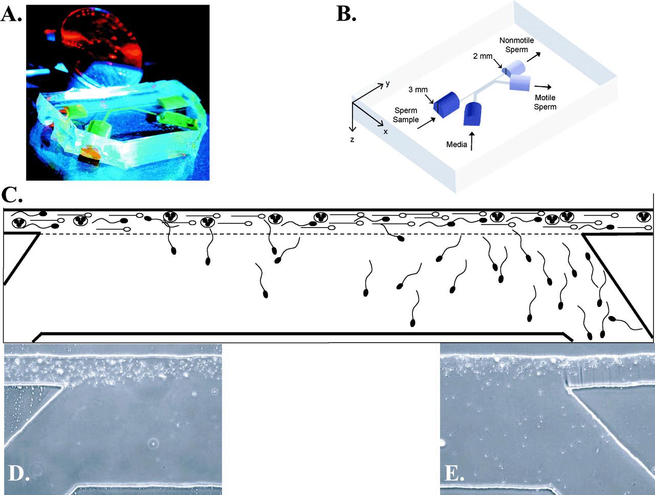
Schuster et al (2003) developed a microfluidic device taking advantage of parallel laminar flow streams present at the microscale. In this device, a flowing stream of semen was placed in parallel with a flowing stream of media within a microchannel. Flow within microchannels was maintained by a novel gravity-driven, horizontally oriented pumping system developed specifically for the device (Cho et al, 2003). As discussed, these 2 parallel laminar flow streams mix only by diffusion. Motile sperm demonstrated the ability to actively propel themselves across contacting surface areas and deviated from the initial streamline into the media stream for collection, whereas nonmotile sperm and cellular debris remained in the initial stream and exited the device (Figure 2).
Testing of this laminar flow sorting system was performed with 40 μL of unprocessed human semen, followed by semen samples artificially filled with debris from a stock solution of round immature germ and white blood cells to simulate poor-quality samples. For unprocessed semen, the device consistently produced a sorted fraction with increased motility (mean 98% motile) and improved Kruger strict sperm morphology (mean 22% normal forms) compared with the initial specimen (mean 44% and 10%, respectively). For debris-filled samples, the device not only concentrated motile sperm (mean 98% motile) within the collected fraction, but was also able to produce a round cell:sperm ratio of 1:33 compared with a 10:1 ratio in the starting specimen (Schuster et al, 2003).
Microfluidics might be particularly suitable for IVF for a number of reasons (Suh et al, 2003). The microenvironment of a microchannel more closely resembles in vivo fertilization conditions than a culture dish or microdrop. Microfluidic channels allow for nonturbulent bathing of gametes with fresh media throughout insemination and coincubation. Sperm-oocyte interactions occur in an active environment, rather than the static conditions present in a culture dish or droplet. In addition, sperm can be predictably delivered via laminar flow to each oocyte within the microchannel, eliminating the randomness of sperm-oocyte interaction. In a culture dish, sperm can travel randomly in any direction, thereby relying on random sperm movement toward the oocytes; however, in a microchannel environment, sperm movement is limited by the direction of flow, allowing for active transport to the oocytes. Finally, microchannel environments use extremely small volumes of media, theoretically requiring fewer sperm to achieve insemination concentrations equal to standard IVF with larger volumes.
Previous investigators have attempted, with some success, to reduce the volume of insemination medium with various low-volume vessels, although none have gained widespread acceptance. Van der Ven et al (1989) tested the use of sterile, nonheparinized hematocrit capillary tubes (75 mm length, 0.9 mm inner diameter) for IVF in humans. Normospermic samples were used with standard culture tubes as controls. Volumes of 5–10 μL containing a range of 500–4000 sperm per oocyte were used in these capillary tubes. Overall fertilization rates between controls and capillary tubes was similar (78% and 66%, respectively), although slightly lower for sperm totals of 500–1000 (56%) compared with 2000–4000 total sperm (79%). Ranoux and Seibel (1990) used embryo cryopreservation straws in volumes up to 200 μL (Ranoux et al, 1988) with 2000–4000 motile sperm. Results compared favorably with controls, with 167 of 322 oocytes (51.8%) fertilized by the straw technique.
We have recently demonstrated that mouse IVF can be conducted successfully within microfluidic channels (unpublished data). Not only are lower total numbers of sperm required because of the use of reduced media volumes, we have also demonstrated murine fertilization within microchannels with lower insemination concentrations, further decreasing sperm requirements. We continue to develop design improvements that will result in increased efficiency and ease of use. Such microfluidic devices should ultimately be useful in clinical IVF, not only for oligospermic patients but potentially as a replacement for standard insemination.
This microfluidic sperm sorting device provided a simple, atraumatic method of obtaining motile sperm of normal morphology from both unprocessed normal semen and poor-quality specimens containing significant debris. A limitation of the device regards the flow rate, estimated at ∼20–40 μL/h. In its current form, it is not capable of processing an entire semen specimen; however, it does provide a means of quickly and easily isolating a small sample of motile sperm of normal morphology for ICSI, insemination in microdrops under oil, or microinsemination in an integrated microfluidic device (Clark et al, 2003; unpublished data). In addition, modifications and improvements in the design are in progress that might allow for large-scale processing in parallel and increased efficiency of flow and sorting (Schuster et al, 2003).
As with any new technology, design plays an important role. Improvements in loading methods, which can allow for visualization under magnification without adjustment of the microscope focus, should improve outcomes. Addition of oocytes and sperm to the device under a microscope, which requires time outside of the humidified 5% CO2 environment, has significant deleterious effects on gamete health and survival. Reduction of this time currently requires a significant learning curve. Last, the implementation of more sophisticated but less operator-reliant mechanisms for fluid flow should improve efficiency. Current studies are focused on a variation of the gravity-driven, horizontally oriented reservoir pumping system (Zhu et al, 2004) from the microfluidic sperm sorter developed by Cho et al (2003) and its application to microfluidic insemination.
Although much of the work with microfluidics in IVF has been performed in a stepwise fashion, the ultimate goal of process miniaturization and microfluidic technology is integration. Use of microfluidic technology for sperm processing ultimately results in a small volume and fraction of motile sperm. Such volumes are difficult to subsequently use and translate into a macroscale environment. However, laminar flow-sorted sperm have been used for subsequent fertilization within a microenvironment (unpublished data). Integration of a microfluidic channel for the oocyte and the collection stream of sorted sperm would result in automatic coincubation of the oocyte with these motile sperm. Following insemination, the oocyte can be directed to a secondary site for cumulus removal, evaluation for fertilization, and embryo culture (see review in Beebe et al, 2002b). Sequential media can be provided for ideal embryo development. Each step logically follows the other, with no cell manipulation other than directing flow along a variety of channels. Miniaturization allows the entire system to be small and self-contained. Decreased intervention by laboratory personnel not only decreases gamete and embryo manipulation but also provides for greater consistency of incubation conditions.
Obviously, multiple hurdles exist in engineering integration of these processes. The development of reliable methods for directing flow through a network of channels is necessary. Recently, numerous active and passive valves (Beebe et al, 2002a) or switches (McDonald et al, 2000) have been designed for microfluidic devices, but the applicability to gamete and embryo manipulation must be demonstrated. Automated delivery of fluid at precise rates is important for sperm sorting, culture media exchange, and embryo manipulation. A passive gravity-driven fluid pump has been employed for microfluidic sperm sorting (Cho et al, 2003), and active hands-on regulation of fluid flow with syringes has been used for insemination and embryo handling (Davis et al, 2000). Recently, we developed a new computer-controlled, integrated, microfluidic control system with up to hundreds of on-chip pumps and valves powered by individually actuated Braille pins on a portable, refreshable Braille display (Figure 3; Gu et al, 2004). Typically these displays are used as a reading tool for the blind. The display can convert electronic signals (from a computer) to vertical translations of 8 small individual pinheads on each of the many Braille cells (typically 8–80 cells for a total of 64–640 individually controlled and actuated pinheads). A line of these cells would typically represent a line of text displayed by a computer. We took advantage of the fast refresh rate and minuscule size of the Braille pinhead by aligning the pinhead's localized pressure onto microchannels, squeezing them shut at high refresh rates. The system takes advantage of the resilient yet elastic nature of PDMS microchannels fabricated with soft lithography and the movement of Braille pins to “squeeze” fluid through channels. Each stroke of a Braille pin can be used to generate a forward or backward flow of liquid through the microchannel when synchronized to various valving patterns. The volume of flow generated per stroke can be controlled by adjusting the volume of liquid displaced by the pin. This method of fluidic control is portable, versatile, and cost effective. Braille displays are commercially available, can be battery powered, and have embedded computerized control in devices the size of a person's hand. With this new concept, we have demonstrated 3 key functions necessary for future application of microfluidics for microinsemination and embryo culture and biochemical analysis system: 1) valve actuation (opening and closing), 2) pumping, and 3) mixing. Finally, methods of fabrication and packaging of microfluidic devices must be refined before widespread acceptance of this technology for human applications. Current devices have been for research purposes. However, development of an IVF laboratory-on-a-chip is a realistic and exciting goal.
也许在过去的30年里,最令人兴奋和革命性的科学发现之一是体外受精(IVF)治疗人类不孕症的发展。自1978年在英格兰的Old-ham首次试管婴儿出生以来,它对许多家庭的影响是无法量化的(Steptoe和Edwards, 1978)。随着辅助生殖技术(ART)的临床应用越来越多,科学家和临床医生对基本的配子和胚胎生物学有了深入的了解,并将这些知识转化为改善体外受精(IVF)的过程。对个别步骤的批判性分析改善了结果。注意精子的处理和分离,以增加活动精子的恢复和减少精子损伤,提高了受精率和胚胎发育(Mortimer, 1994)。使用胞浆内单精子注射(ICSI)即使在精子质量或数量严重受损的情况下也可以受精(Bonduelle等,1999)。最后,胚胎培养的改进导致体外胚胎发育和着床率的提高(Gardner和Lane, 1998;池,2002)。然而,大多数科学注意力集中在方法上,而不是技术开发和设备上。无论采用何种技术,精液仍然在试管中进行处理,精子在处理后与卵母细胞一起物理放置,受精和胚胎培养在培养皿、试管或两者中进行,且体积相对较大(Trounson and Gardner, 2000)。除了配子和胚胎显微操作外,体外受精的技术进步尚未得到广泛应用。然而,正是这些技术的进步,而不是程序或方法的改变,对辅助生殖产生了最大的影响。微流体技术是一种很有前途的新技术,它的存在和研究日益深入。这项技术有望成为体外受精过程中每一步的替代方案。微流体学基于微环境中流体行为的物理原理,已广泛应用于化学和分子生物学领域(Tomlinson et al ., 1995)。目前,微流体学在细胞行为和相互作用的研究中越来越受到关注(Shim et al ., 2003)。在本文中,我们介绍了微观尺度下流体行为的基础知识,并重点介绍了该技术在生殖科学之外的先前应用。然后,我们描述了设备的制造,并回顾了在精子分选和微授精中使用微流体的初步研究。最后,指出了微流控技术的局限性,并对微流控技术在ART中的应用前景和发展方向进行了展望。流体力学是一门复杂的物理和数学科学;因此,流体物理的广泛技术描述和审查超出了本审查的范围和意图。相反,将讨论控制微环境中流体行为的基本原则,特别是那些与目前正在开发的试管婴儿设备和技术有特定联系的方面。我们有意避免包括数学细节,而是选择传达微通道中流体力学的一般概念。Beebe et al . (2002a)和Brody et al .(1996)对微流控物理进行了全面的技术和数学描述。在微观尺度上,流体受到的力在我们日常生活中通常是不重要的。在我们正常环境的尺度上,流体是动荡的;流体中的粒子以一种不可预测的方式运动。紊流取决于某些流体特性(粘度、密度和速度)以及通道的几何形状和大小,从而计算出一个称为雷诺数的值。当通道的尺度达到微米级时,雷诺数减少,并且越来越依赖于流体特性。当雷诺数低于某一阈值时,流体以层流方式流动。简而言之,微通道内的流动变得流线型和可预测(图1)。在微观尺度上,流体行为越来越受到粘性力和表面张力的控制,这可以被描述为液体分子的内聚性。粘性力的这种主导作用导致了几个有趣的现象。低雷诺数的流几乎没有动量;因此,微通道内的流体对外力的变化作出快速而可靠的反应。此外,在微观尺度上,相互接触的两股或更多的层流不会混合,除非分子在层流的界面上扩散。在微观尺度上,接触面之间的扩散速度可以非常快,部分原因是跨越流体体积所需的距离相对较短。 许多这些流体特性在微尺度形成的原则驱动的兴趣使用微通道配子和胚胎操作。一般来说,微环境与培养皿或培养基相比,更接近于体内受精和发育的条件。下面,我们将讨论研究微流体在男科中的应用背后的理论,它的测试、限制和潜在的未来影响。对微流体的兴趣始于实验室中化学和生物分析装置小型化的尝试(Kricka, 1998年)。目前的设计通常被称为“芯片上的实验室”或微总量分析系统(μTAS),其功能是允许流体在其微型通道和腔室内流动时发生各种化学过程和相互作用(Weigl和Yager, 1999)。这些设备执行其目的所需的所有分析功能,包括样品处理,混合,孵育,分选,运输,相互作用,以及在集成的微流控“芯片”内检测或信号。例子包括,但不限于,血清中抗体的免疫测定(Linder等人,2002)和酶反应动力学测定(Xue等人,2001;Yakovleva et al, 2002)。细胞生物学中的其他应用已经出现,例如在微观尺度上工作的集成细胞分选装置(Fu等人,2002)和允许研究细胞与底物或其他细胞相互作用的微流体装置(Shim等人,2003)。利用微流体和层流原理证明了细胞生物学的进步,允许选择性地将感兴趣的亚细胞区域暴露于膜渗透分子(Takayama等人,2001年)。如此精确地将分子传递到细胞子域,说明了微流体调节流体流动的精度。这种芯片实验室技术的优势是多方面的。首先,一旦设计和测试,这种设备的制造是直接和廉价的,允许它们是一次性的(McDonald等人,2000)。微流控分析设备使用非常小体积的样品和试剂,提供更快的反应和响应时间(Weigl和Yager, 1999)。小型化非常重要,它允许在一个小的、自包含的单元内集成多个过程(Kricka, 1998)。这可以转换为多个并行分析,连续串行过程,或两者兼而有之。这里给出的简要概述只是为了让读者熟悉微流控技术的各种功能,而绝不是微流控在科学中的应用的全面列表。鼓励读者查阅更全面的评论(Khandurina and Guttman, 2002;Verpoorte, 2002)。微流体系统最初是用工业中常见的材料和技术制造出来的——微电子(McDonald et al ., 2000)。硅和玻璃的光刻和蚀刻是一项高度发达的技术,对小型化分析系统感兴趣的研究人员也很容易获得,但成本是一个重大障碍。为了寻找合适的替代品,聚合物迅速成为微流体生物装置制造的材料(McDonald等人,2000)。诸如聚甲基丙烯酸甲酯(Martynova等人,1997年)、氟化乙丙烯(Sahlin等人,2002年)和聚二甲基硅氧烷(PDMS;McDonald和Whitesides, 2002)比硅玻璃替代品更便宜,更容易操作(Martynova等人,1997)。特别是PDMS已成为迄今为止最积极探索和最有前途的材料之一,具有许多特别适合生物用途的特性。它是无毒的,透明的,绝缘的,可渗透的气体(麦克唐纳和怀特塞德斯,2002年)。从制造的角度来看,PDMS可以实现亚微米级的成型保真度,在低温下固化,并且可以很容易地对自身和许多其他材料进行可逆密封(McDonald等,2000)。虽然PDMS通常被认为是无毒的,但必须特别考虑它与配子和胚胎的使用,与转化细胞系相比,配子和胚胎对环境非常敏感。在使用精子微流体装置之前,测试证实,长时间暴露在制造过程中使用的材料中不会产生负面影响。Schuster等人(2003)报道,暴露于PDMS 30分钟不会改变精子存活率。此外,格拉斯哥等人(2001)发现,与对照组相比,连续暴露于大量光印化合物中,2细胞小鼠胚胎向囊胚期的发育没有发生变化。因此,pdms组成的微通道或用于其构建的材料似乎不会对配子或胚胎产生有害影响。 最后,微通道环境使用非常小体积的培养基,理论上需要更少的精子来达到与大体积标准试管婴儿相同的授精浓度。以前的研究人员曾尝试用各种小容量的试管来减少受精介质的体积,并取得了一些成功,尽管没有一种方法得到广泛的接受。Van der Ven等人(1989)测试了无菌、非肝素化的红细胞压层毛细血管(75毫米长,0.9毫米内径)在人类试管婴儿中的应用。正常精子标本以标准培养管为对照。在这些毛细管中使用5-10 μL的体积,每个卵母细胞含有500-4000个精子。对照组和毛细管组的总体受精率相似(分别为78%和66%),尽管500-1000个精子总数(56%)略低于2000-4000个精子总数(79%)。Ranoux和Seibel(1990)使用容量达200 μL的胚胎低温保存吸管(Ranoux et al ., 1988)保存2000-4000个活动精子。结果与对照组相比,322个卵母细胞中有167个(51.8%)通过秸秆技术受精。我们最近证明,小鼠体外受精可以在微流体通道内成功进行(未发表的数据)。由于使用较少的培养基体积,不仅需要较少的精子总数,而且我们还证明了小鼠在微通道内受精,其授精浓度较低,进一步降低了精子需求。我们将继续开发设计改进,以提高效率和易用性。这种微流体装置最终将在临床试管婴儿中发挥作用,不仅适用于少精子患者,而且有可能替代标准的人工授精。这种微流控精子分选装置提供了一种简单、无损伤的方法,可以从未处理的正常精液和含有大量碎片的低质量样本中获得正常形态的活动精子。该装置的一个限制是流速,估计为~ 20-40 μL/h。以目前的形式,它无法处理整个精液标本;然而,它确实提供了一种快速、方便地分离少量正常形态的活动精子样本的方法,用于ICSI、油下微滴人工授精或集成微流体装置中的微授精(Clark et al ., 2003;未公开的数据)。此外,设计上的修改和改进正在进行中,这可能会允许大规模的并行处理,并提高流动和分类的效率(Schuster等人,2003)。与任何新技术一样,设计扮演着重要的角色。加载方法的改进,可以允许在放大镜下可视化而无需调整显微镜焦点,应该改善结果。在显微镜下将卵母细胞和精子添加到设备中,这需要在5%二氧化碳的潮湿环境外的时间,对配子的健康和生存有显著的有害影响。减少这个时间目前需要一个重要的学习曲线。最后,采用更复杂但对作业者较少依赖的流体流动机制应该能提高效率。目前的研究主要集中在Cho等人(2003)开发的微流控精子分选器的重力驱动、水平导向的储层泵送系统(Zhu等,2004)的变体及其在微流控人工授精中的应用。尽管体外受精中微流控技术的大部分工作都是以循序渐进的方式进行的,但过程小型化和微流控技术的最终目标是集成。使用微流控技术进行精子处理,最终产生小体积和一小部分可运动精子。这样的体量很难随后使用并转化为宏观尺度的环境。然而,层流分类的精子已被用于微环境中的后续受精(未发表的数据)。卵母细胞的微流控通道和已分类精子的收集流的集成将导致卵母细胞与这些活动精子的自动共孵育。授精后,卵母细胞可以被引导到第二个部位进行积云清除、受精评估和胚胎培养(见Beebe et al, 2002b的综述)。序贯培养基可为理想的胚胎发育提供条件。每个步骤在逻辑上遵循另一个步骤,除了沿着各种通道引导流动之外,没有细胞操作。小型化使整个系统变得小而独立。减少实验室人员的干预不仅减少了配子和胚胎的操作,而且提供了更大的孵育条件一致性。显然,在这些过程的工程集成中存在多个障碍。开发可靠的方法来引导水流通过管道网络是必要的。


 分享
分享
 求助内容:
求助内容: 应助结果提醒方式:
 扫码关注我们
