{"title":"Relation between lipogranuloma formation and fibrosis, and the origin of brown pigments in lipogranuloma of the canine liver.","authors":"Kaori Isobe, Hiroyuki Nakayama, Koji Uetsuka","doi":"10.1186/1476-5926-7-5","DOIUrl":null,"url":null,"abstract":"<p><strong>Background: </strong>In a previous study we confirmed that canine hepatic lipogranuloma, defined as lesions consisting of small round cells which contain lipid vacuoles and brown pigments in their cytoplasm, was an assembly of Kupffer cells and/or macrophages, and that the cytoplasmic brown pigments in the lesions were hemosiderin and ceroid. However, the pathogenesis of the lesion remains unclear. Kupffer cells (resident macrophages) play a key role in hepatic fibrogenesis due to the production of cytokines including TGF-beta. In the present study, we have examined 52 canine liver samples (age: newborn - 14 years; 25 males and 27 females) and investigated the correlation between lipogranuloma formation and fibrosis as well as the origin of brown pigments of lipogranulomas.</p><p><strong>Results: </strong>Lipogranulomas were detected histopathologically in 23 (44.2%) of the 52 liver samples. No significant correlation was found between the density of lipogranulomas and distribution of collagen type I/III in the liver. Pigmentation of lipogranulomas showed significant correlations with that on both hepatocytes and sinusoidal cells, indicating that pigments of lipogranuloma (hemosiderin and ceroid) might be derived from hepatocytes and Kupffer cells.</p><p><strong>Conclusion: </strong>Lipogranulomas are not a contributing factor in hepatic fibrosis, but might be a potential indicator of the accumulation of iron and lipid inside the liver.</p>","PeriodicalId":84474,"journal":{"name":"Comparative hepatology","volume":"7 ","pages":"5"},"PeriodicalIF":0.0000,"publicationDate":"2008-05-12","publicationTypes":"Journal Article","fieldsOfStudy":null,"isOpenAccess":false,"openAccessPdf":"https://sci-hub-pdf.com/10.1186/1476-5926-7-5","citationCount":"11","resultStr":null,"platform":"Semanticscholar","paperid":null,"PeriodicalName":"Comparative hepatology","FirstCategoryId":"1085","ListUrlMain":"https://doi.org/10.1186/1476-5926-7-5","RegionNum":0,"RegionCategory":null,"ArticlePicture":[],"TitleCN":null,"AbstractTextCN":null,"PMCID":null,"EPubDate":"","PubModel":"","JCR":"","JCRName":"","Score":null,"Total":0}
引用次数: 11
Abstract
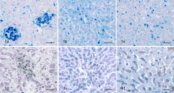
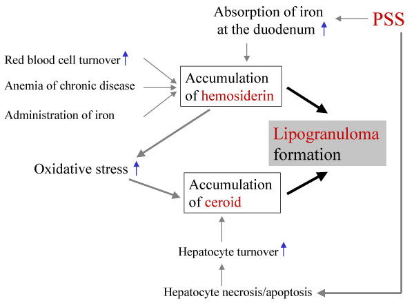
Background: In a previous study we confirmed that canine hepatic lipogranuloma, defined as lesions consisting of small round cells which contain lipid vacuoles and brown pigments in their cytoplasm, was an assembly of Kupffer cells and/or macrophages, and that the cytoplasmic brown pigments in the lesions were hemosiderin and ceroid. However, the pathogenesis of the lesion remains unclear. Kupffer cells (resident macrophages) play a key role in hepatic fibrogenesis due to the production of cytokines including TGF-beta. In the present study, we have examined 52 canine liver samples (age: newborn - 14 years; 25 males and 27 females) and investigated the correlation between lipogranuloma formation and fibrosis as well as the origin of brown pigments of lipogranulomas.
Results: Lipogranulomas were detected histopathologically in 23 (44.2%) of the 52 liver samples. No significant correlation was found between the density of lipogranulomas and distribution of collagen type I/III in the liver. Pigmentation of lipogranulomas showed significant correlations with that on both hepatocytes and sinusoidal cells, indicating that pigments of lipogranuloma (hemosiderin and ceroid) might be derived from hepatocytes and Kupffer cells.
Conclusion: Lipogranulomas are not a contributing factor in hepatic fibrosis, but might be a potential indicator of the accumulation of iron and lipid inside the liver.


 分享
分享

 求助内容:
求助内容: 应助结果提醒方式:
 扫码关注我们
