{"title":"Non invasive in vivo investigation of hepatobiliary structure and function in STII medaka (Oryzias latipes): methodology and applications.","authors":"Ron C Hardman, Seth W Kullman, David E Hinton","doi":"10.1186/1476-5926-7-7","DOIUrl":null,"url":null,"abstract":"<p><strong>Background: </strong>A novel transparent stock of medaka (Oryzias latipes; STII), recessive for all pigments found in chromatophores, permits transcutaneous imaging of internal organs and tissues in living individuals. Findings presented describe the development of methodologies for non invasive in vivo investigation in STII medaka, and the successful application of these methodologies to in vivo study of hepatobiliary structure, function, and xenobiotic response, in both 2 and 3 dimensions.</p><p><strong>Results: </strong>Using brightfield, and widefield and confocal fluorescence microscopy, coupled with the in vivo application of fluorescent probes, structural and functional features of the hepatobiliary system, and xenobiotic induced toxicity, were imaged at the cellular level, with high resolution (< 1 microm), in living individuals. The findings presented demonstrate; (1) phenotypic response to xenobiotic exposure can be investigated/imaged in vivo with high resolution (< 1 microm), (2) hepatobiliary transport of solutes from blood to bile can be qualitatively and quantitatively studied/imaged in vivo, (3) hepatobiliary architecture in this lower vertebrate liver can be studied in 3 dimensions, and (4) non invasive in vivo imaging/description of hepatobiliary development in this model can be investigated.</p><p><strong>Conclusion: </strong>The non-invasive in vivo methodologies described are a unique means by which to investigate biological structure, function and xenobiotic response with high resolution in STII medaka. In vivo methodologies also provide the future opportunity to integrate molecular mechanisms (e.g., genomic, proteomic) of disease and toxicity with phenotypic changes at the cellular and system levels of biological organization. While our focus has been the hepatobiliary system, other organ systems are equally amenable to in vivo study, and we consider the potential for discovery, within the context of in vivo investigation in STII medaka, as significant.</p>","PeriodicalId":84474,"journal":{"name":"Comparative hepatology","volume":"7 ","pages":"7"},"PeriodicalIF":0.0000,"publicationDate":"2008-10-06","publicationTypes":"Journal Article","fieldsOfStudy":null,"isOpenAccess":false,"openAccessPdf":"https://sci-hub-pdf.com/10.1186/1476-5926-7-7","citationCount":"4","resultStr":null,"platform":"Semanticscholar","paperid":null,"PeriodicalName":"Comparative hepatology","FirstCategoryId":"1085","ListUrlMain":"https://doi.org/10.1186/1476-5926-7-7","RegionNum":0,"RegionCategory":null,"ArticlePicture":[],"TitleCN":null,"AbstractTextCN":null,"PMCID":null,"EPubDate":"","PubModel":"","JCR":"","JCRName":"","Score":null,"Total":0}
引用次数: 4
Abstract
Background: A novel transparent stock of medaka (Oryzias latipes; STII), recessive for all pigments found in chromatophores, permits transcutaneous imaging of internal organs and tissues in living individuals. Findings presented describe the development of methodologies for non invasive in vivo investigation in STII medaka, and the successful application of these methodologies to in vivo study of hepatobiliary structure, function, and xenobiotic response, in both 2 and 3 dimensions.
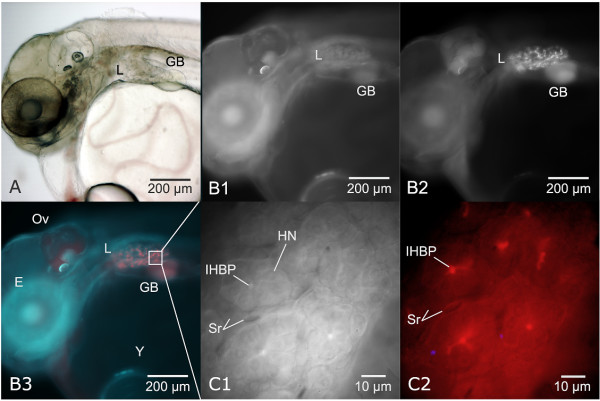
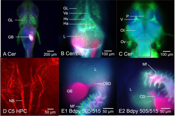
Results: Using brightfield, and widefield and confocal fluorescence microscopy, coupled with the in vivo application of fluorescent probes, structural and functional features of the hepatobiliary system, and xenobiotic induced toxicity, were imaged at the cellular level, with high resolution (< 1 microm), in living individuals. The findings presented demonstrate; (1) phenotypic response to xenobiotic exposure can be investigated/imaged in vivo with high resolution (< 1 microm), (2) hepatobiliary transport of solutes from blood to bile can be qualitatively and quantitatively studied/imaged in vivo, (3) hepatobiliary architecture in this lower vertebrate liver can be studied in 3 dimensions, and (4) non invasive in vivo imaging/description of hepatobiliary development in this model can be investigated.
Conclusion: The non-invasive in vivo methodologies described are a unique means by which to investigate biological structure, function and xenobiotic response with high resolution in STII medaka. In vivo methodologies also provide the future opportunity to integrate molecular mechanisms (e.g., genomic, proteomic) of disease and toxicity with phenotypic changes at the cellular and system levels of biological organization. While our focus has been the hepatobiliary system, other organ systems are equally amenable to in vivo study, and we consider the potential for discovery, within the context of in vivo investigation in STII medaka, as significant.


 分享
分享

 求助内容:
求助内容: 应助结果提醒方式:
 扫码关注我们
