Renee G H M van Sprundel, Ted S G A M van den Ingh, Valeer J Desmet, Azeam Katoonizadeh, Louis C Penning, Jan Rothuizen, Tania Roskams, Bart Spee
{"title":"Keratin 19 marks poor differentiation and a more aggressive behaviour in canine and human hepatocellular tumours.","authors":"Renee G H M van Sprundel, Ted S G A M van den Ingh, Valeer J Desmet, Azeam Katoonizadeh, Louis C Penning, Jan Rothuizen, Tania Roskams, Bart Spee","doi":"10.1186/1476-5926-9-4","DOIUrl":null,"url":null,"abstract":"<p><strong>Background: </strong>The expression of Keratin 19 (K19) was reported in a subset of hepatocellular carcinomas (HCCs). K19 positive HCCs are associated with an increased malignancy compared to K19 negative HCCs. No suitable mouse models exist for this subtype of HCC, nor is the incidence of K19 expression in hepatocellular neoplasia in model animals known. Therefore, we compared the occurrence and tumour behaviour of K19 positive hepatocellular neoplasias in dog and man.</p><p><strong>Results: </strong>The expression of hepatocellular differentiation (HepPar-1), biliary/progenitor cell (K7, K19), and malignancy (glypican-3) markers was semi-quantitatively assessed by immunohistochemistry. The histological grade of tumour differentiation was determined according to a modified classification of Edmondson and Steiner; the staging included intrahepatic, lymph node or distant metastases. Four of the 34 canine hepatocellular neoplasias showed K19 positivity (12%), of which two co-expressed K7. K19 positive tumours did not express HepPar-1, despite the histological evidence of a hepatocellular origin. Like in human HCC, all K19 positive hepatocellular neoplasias were glypican-3 positive and histologically poorly differentiated and revealed intra- or extrahepatic metastases whereas K19 negative hepatocellular neoplasias did not.</p><p><strong>Conclusions: </strong>K19 positive hepatocellular neoplasias are highly comparable to man and occur in 12% of canine hepatocellular tumours and are associated with a poorly differentiated histology and aggressive tumour behaviour.</p>","PeriodicalId":84474,"journal":{"name":"Comparative hepatology","volume":"9 1","pages":"4"},"PeriodicalIF":0.0000,"publicationDate":"2010-02-18","publicationTypes":"Journal Article","fieldsOfStudy":null,"isOpenAccess":false,"openAccessPdf":"https://sci-hub-pdf.com/10.1186/1476-5926-9-4","citationCount":"34","resultStr":null,"platform":"Semanticscholar","paperid":null,"PeriodicalName":"Comparative hepatology","FirstCategoryId":"1085","ListUrlMain":"https://doi.org/10.1186/1476-5926-9-4","RegionNum":0,"RegionCategory":null,"ArticlePicture":[],"TitleCN":null,"AbstractTextCN":null,"PMCID":null,"EPubDate":"","PubModel":"","JCR":"","JCRName":"","Score":null,"Total":0}
引用次数: 34
Abstract
Background: The expression of Keratin 19 (K19) was reported in a subset of hepatocellular carcinomas (HCCs). K19 positive HCCs are associated with an increased malignancy compared to K19 negative HCCs. No suitable mouse models exist for this subtype of HCC, nor is the incidence of K19 expression in hepatocellular neoplasia in model animals known. Therefore, we compared the occurrence and tumour behaviour of K19 positive hepatocellular neoplasias in dog and man.
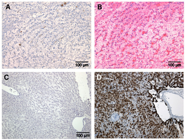
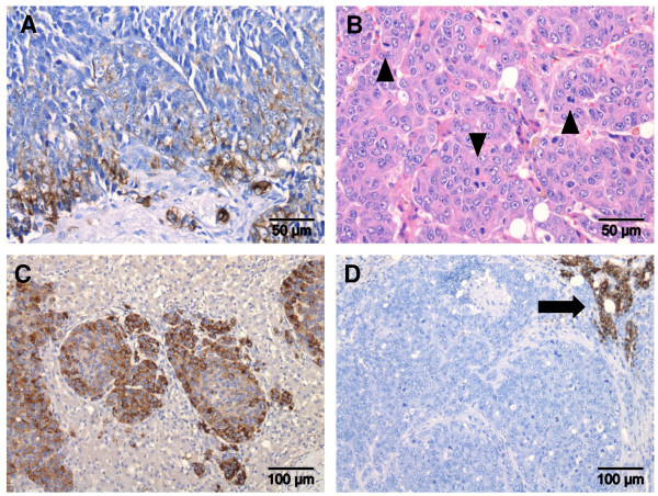
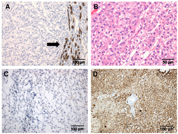
Results: The expression of hepatocellular differentiation (HepPar-1), biliary/progenitor cell (K7, K19), and malignancy (glypican-3) markers was semi-quantitatively assessed by immunohistochemistry. The histological grade of tumour differentiation was determined according to a modified classification of Edmondson and Steiner; the staging included intrahepatic, lymph node or distant metastases. Four of the 34 canine hepatocellular neoplasias showed K19 positivity (12%), of which two co-expressed K7. K19 positive tumours did not express HepPar-1, despite the histological evidence of a hepatocellular origin. Like in human HCC, all K19 positive hepatocellular neoplasias were glypican-3 positive and histologically poorly differentiated and revealed intra- or extrahepatic metastases whereas K19 negative hepatocellular neoplasias did not.
Conclusions: K19 positive hepatocellular neoplasias are highly comparable to man and occur in 12% of canine hepatocellular tumours and are associated with a poorly differentiated histology and aggressive tumour behaviour.


 分享
分享

 求助内容:
求助内容: 应助结果提醒方式:
 扫码关注我们
