Hee Won Moon, Tae Young Kim, Bo Ra Oh, Sang Mee Hwang, Jiseok Kwon, Ja-Lok Ku, Dong Soon Lee
{"title":"Effects of granulocyte-colony stimulating factor and the expression of its receptor on various malignant cells.","authors":"Hee Won Moon, Tae Young Kim, Bo Ra Oh, Sang Mee Hwang, Jiseok Kwon, Ja-Lok Ku, Dong Soon Lee","doi":"10.5045/kjh.2012.47.3.219","DOIUrl":null,"url":null,"abstract":"<p><strong>Background: </strong>Granulocyte-colony stimulating factor (G-CSF) is extensively used to improve neutrophil count during anti-cancer chemotherapy. We investigated the effects of G-CSF on several leukemic cell lines and screened for the expression of the G-CSF receptor (G-CSFR) in various malignant cells.</p><p><strong>Methods: </strong>We examined the effects of the most commonly used commercial forms of G-CSF (glycosylated lenograstim and nonglycosylated filgrastim) on various leukemic cell lines by flow cytometry. Moreover, we screened for the expression of G-CSFR mRNA in 38 solid tumor cell lines by using real-time PCR.</p><p><strong>Results: </strong>G-CSF stimulated proliferation (40-80% increase in proliferation in treated cells as compared to that in control cells) in 3 leukemic cell lines and induced differentiation of AML1/ETO+ leukemic cells. Among the 38 solid tumor cell lines, 5 cell lines (hepatoblastoma, 2 breast carcinoma, squamous cell carcinoma of the larynx, and melanoma cell lines) showed G-CSFR mRNA expression.</p><p><strong>Conclusion: </strong>The results of the present study show that therapeutic G-CSF might stimulate the proliferation and differentiation of malignant cells with G-CSFR expression, suggesting that prescreening for G-CSFR expression in primary tumor cells may be necessary before using G-CSF for treatment.</p>","PeriodicalId":23001,"journal":{"name":"The Korean Journal of Hematology","volume":"47 3","pages":"219-24"},"PeriodicalIF":0.0000,"publicationDate":"2012-09-01","publicationTypes":"Journal Article","fieldsOfStudy":null,"isOpenAccess":false,"openAccessPdf":"https://sci-hub-pdf.com/10.5045/kjh.2012.47.3.219","citationCount":"7","resultStr":null,"platform":"Semanticscholar","paperid":null,"PeriodicalName":"The Korean Journal of Hematology","FirstCategoryId":"1085","ListUrlMain":"https://doi.org/10.5045/kjh.2012.47.3.219","RegionNum":0,"RegionCategory":null,"ArticlePicture":[],"TitleCN":null,"AbstractTextCN":null,"PMCID":null,"EPubDate":"2012/9/25 0:00:00","PubModel":"Epub","JCR":"","JCRName":"","Score":null,"Total":0}
引用次数: 7
Abstract
Background: Granulocyte-colony stimulating factor (G-CSF) is extensively used to improve neutrophil count during anti-cancer chemotherapy. We investigated the effects of G-CSF on several leukemic cell lines and screened for the expression of the G-CSF receptor (G-CSFR) in various malignant cells.
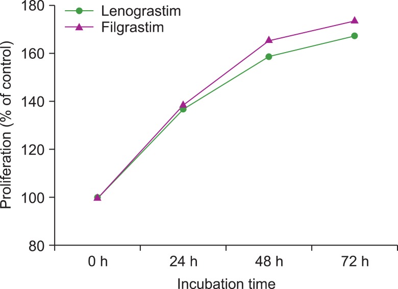
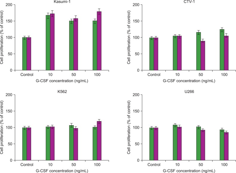
Methods: We examined the effects of the most commonly used commercial forms of G-CSF (glycosylated lenograstim and nonglycosylated filgrastim) on various leukemic cell lines by flow cytometry. Moreover, we screened for the expression of G-CSFR mRNA in 38 solid tumor cell lines by using real-time PCR.
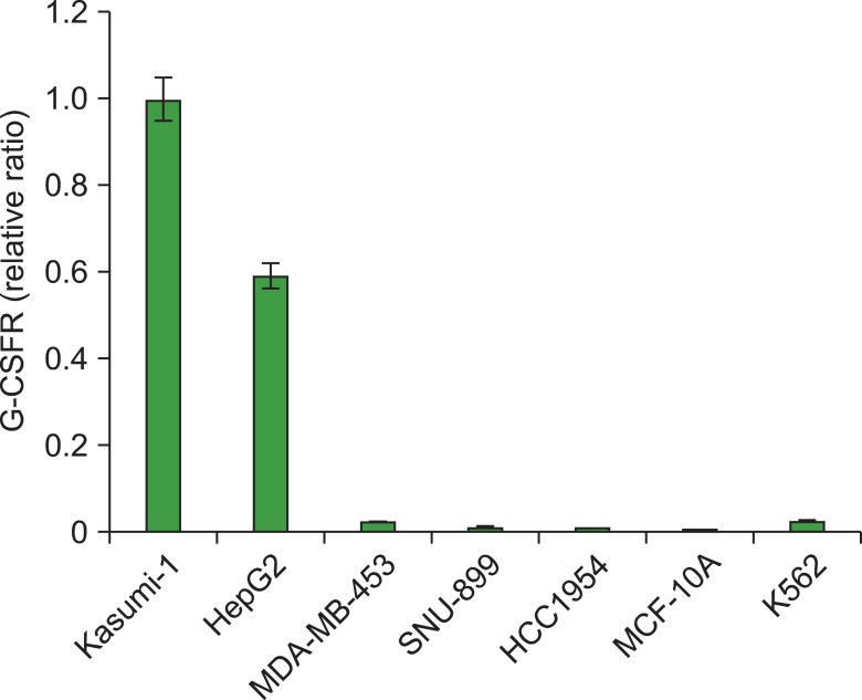
Results: G-CSF stimulated proliferation (40-80% increase in proliferation in treated cells as compared to that in control cells) in 3 leukemic cell lines and induced differentiation of AML1/ETO+ leukemic cells. Among the 38 solid tumor cell lines, 5 cell lines (hepatoblastoma, 2 breast carcinoma, squamous cell carcinoma of the larynx, and melanoma cell lines) showed G-CSFR mRNA expression.
Conclusion: The results of the present study show that therapeutic G-CSF might stimulate the proliferation and differentiation of malignant cells with G-CSFR expression, suggesting that prescreening for G-CSFR expression in primary tumor cells may be necessary before using G-CSF for treatment.


 分享
分享

 求助内容:
求助内容: 应助结果提醒方式:
 扫码关注我们
