{"title":"Tannic acid label indicates abnormal cell development coinciding with regeneration of renal tubules.","authors":"Will W Minuth, Lucia Denk","doi":"10.1186/1472-6890-14-34","DOIUrl":null,"url":null,"abstract":"<p><strong>Background: </strong>Stem/progenitor cells are in the focus of research as a future therapeutic option to stimulate regeneration in diseased renal parenchyma. However, current data indicate that successful seeding of implanted stem/progenitor cells is prevented by harmful interstitial fluid and altered extracellular matrix. To find out possible parameters for cell adaptation, the present investigation was performed.</p><p><strong>Methods: </strong>Renal stem/progenitor cells were mounted in an artificial interstitium for perfusion culture. Exposure to chemically defined but CO2-independent culture media was tested during 13 days. Cell biological features were then analyzed by histochemistry, while structural details were investigated by transmission electron microscopy after conventional and improved fixation of specimens.</p><p><strong>Results: </strong>Culture of renal stem/progenitor cells as well in Leibovitz's L-15 Medium as CO2 Independent Medium shows in fluorescence microscopy spatial development of numerous tubules. Specimens of both media fixed by conventional glutaraldehyde exhibit in electron microscopy a homogeneous cell population in developed tubules. In contrast, fixation by glutaraldehyde including tannic acid illuminates that dispersed dark marked cells of unknown function are present. The screening further demonstrates that the dark cell type does not comply with cells found in embryonic, maturing or matured renal parenchyma.</p><p><strong>Conclusions: </strong>The actual data show that development of abnormal cell features must be taken into account, when regeneration of renal tubules is simulated under in vitro conditions.</p>","PeriodicalId":35804,"journal":{"name":"BMC Clinical Pathology","volume":"14 ","pages":"34"},"PeriodicalIF":0.0000,"publicationDate":"2014-07-15","publicationTypes":"Journal Article","fieldsOfStudy":null,"isOpenAccess":false,"openAccessPdf":"https://sci-hub-pdf.com/10.1186/1472-6890-14-34","citationCount":"5","resultStr":null,"platform":"Semanticscholar","paperid":null,"PeriodicalName":"BMC Clinical Pathology","FirstCategoryId":"1085","ListUrlMain":"https://doi.org/10.1186/1472-6890-14-34","RegionNum":0,"RegionCategory":null,"ArticlePicture":[],"TitleCN":null,"AbstractTextCN":null,"PMCID":null,"EPubDate":"2014/1/1 0:00:00","PubModel":"eCollection","JCR":"Q2","JCRName":"Medicine","Score":null,"Total":0}
引用次数: 5
Abstract
Background: Stem/progenitor cells are in the focus of research as a future therapeutic option to stimulate regeneration in diseased renal parenchyma. However, current data indicate that successful seeding of implanted stem/progenitor cells is prevented by harmful interstitial fluid and altered extracellular matrix. To find out possible parameters for cell adaptation, the present investigation was performed.
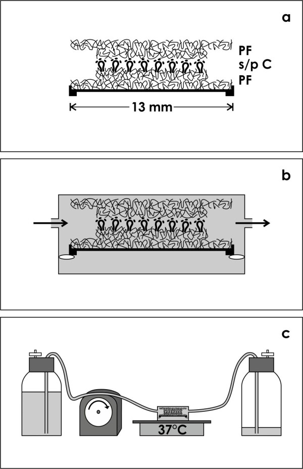
Methods: Renal stem/progenitor cells were mounted in an artificial interstitium for perfusion culture. Exposure to chemically defined but CO2-independent culture media was tested during 13 days. Cell biological features were then analyzed by histochemistry, while structural details were investigated by transmission electron microscopy after conventional and improved fixation of specimens.
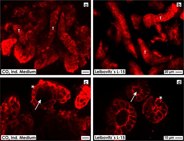
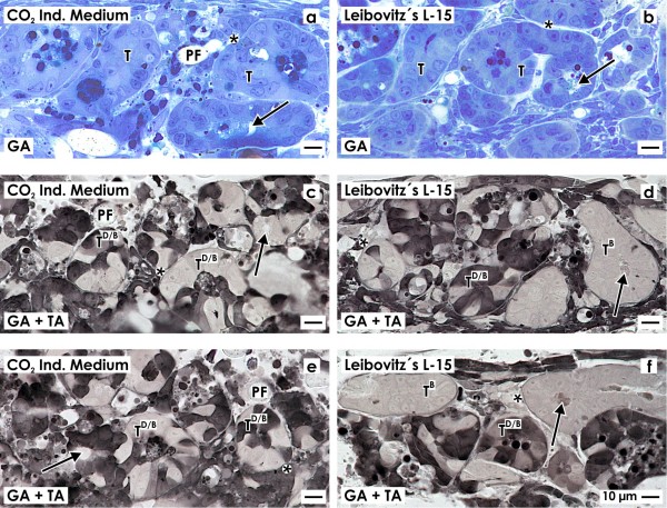
Results: Culture of renal stem/progenitor cells as well in Leibovitz's L-15 Medium as CO2 Independent Medium shows in fluorescence microscopy spatial development of numerous tubules. Specimens of both media fixed by conventional glutaraldehyde exhibit in electron microscopy a homogeneous cell population in developed tubules. In contrast, fixation by glutaraldehyde including tannic acid illuminates that dispersed dark marked cells of unknown function are present. The screening further demonstrates that the dark cell type does not comply with cells found in embryonic, maturing or matured renal parenchyma.
Conclusions: The actual data show that development of abnormal cell features must be taken into account, when regeneration of renal tubules is simulated under in vitro conditions.
期刊介绍:
BMC Clinical Pathology is an open access journal publishing original peer-reviewed research articles in all aspects of histopathology, haematology, clinical biochemistry, and medical microbiology (including virology, parasitology, and infection control). BMC Clinical Pathology (ISSN 1472-6890) is indexed/tracked/covered by PubMed, CAS, EMBASE, Scopus and Google Scholar.


 分享
分享
 求助内容:
求助内容: 应助结果提醒方式:
 扫码关注我们
