Alessando Lamira, Jardel Francisco Mazzi-Chaves, Laura Ferreira Pinheiro Nicolielo, Graziela Bianchi Leoni, Alice Corrêa Silva-Sousa, Yara Terezinha Corrêa Silva-Sousa, Ruben Pauwels, Nico Buls, Reinhilde Jacobs, Manoel Damião Sousa-Neto
{"title":"CBCT-based assessment of root canal treatment using micro-CT reference images.","authors":"Alessando Lamira, Jardel Francisco Mazzi-Chaves, Laura Ferreira Pinheiro Nicolielo, Graziela Bianchi Leoni, Alice Corrêa Silva-Sousa, Yara Terezinha Corrêa Silva-Sousa, Ruben Pauwels, Nico Buls, Reinhilde Jacobs, Manoel Damião Sousa-Neto","doi":"10.5624/isd.20220019","DOIUrl":null,"url":null,"abstract":"<p><strong>Purpose: </strong>This study compared the root canal anatomy between cone-beam computed tomography (CBCT) and micro-computed tomography (micro-CT) images before and after biomechanical preparation and root canal filling.</p><p><strong>Materials and methods: </strong>Isthmus-containing mesial roots of mandibular molars (n=14) were scanned by micro-CT and 3 CBCT devices: 3D Accuitomo 170 (ACC), NewTom 5G (N5G) and NewTom VGi evo (NEVO). Two calibrated observers evaluated the images for 2-dimensional quantitative parameters, the presence of debris or root perforation, and filling quality in the root canal and isthmus. The kappa coefficient, analysis of variance, and the Tukey test were used for statistical analyses (α=5%).</p><p><strong>Results: </strong>Substantial intra-observer agreement (κ=0.63) was found between micro-CT and ACC, N5G, and NEVO. Debris detection was difficult using ACC (42.9%), N5G (40.0%), and NEVO (40%), with no agreement between micro-CT and ACC, N5G, and NEVO (0.05<κ<0.12). After biomechanical preparation, 2.4%-4.8% of CBCT images showed root perforation that was absent on micro-CT. The 2D parameters showed satisfactory reproducibility between micro-CT and ACC, N5G, and NEVO (intraclass correlation coefficient: 0.60-0.73). Partially filled isthmuses were observed in 2.9% of the ACC images, 8.8% of the N5G and NEVO images, and 26.5% of the micro-CT images, with no agreement between micro-CT and ACC, and poor agreement between micro-CT and N5G and NEVO. Excellent agreement was found for area, perimeter, and the major and minor diameters, while the roundness measures were satisfactory.</p><p><strong>Conclusion: </strong>CBCT images aided in isthmus detection and classification, but did not allow their classification after biomechanical preparation and root canal filling.</p>","PeriodicalId":51714,"journal":{"name":"Imaging Science in Dentistry","volume":"52 3","pages":"245-258"},"PeriodicalIF":2.1000,"publicationDate":"2022-09-01","publicationTypes":"Journal Article","fieldsOfStudy":null,"isOpenAccess":false,"openAccessPdf":"https://ftp.ncbi.nlm.nih.gov/pub/pmc/oa_pdf/52/0a/isd-52-245.PMC9530298.pdf","citationCount":"0","resultStr":null,"platform":"Semanticscholar","paperid":null,"PeriodicalName":"Imaging Science in Dentistry","FirstCategoryId":"1085","ListUrlMain":"https://doi.org/10.5624/isd.20220019","RegionNum":0,"RegionCategory":null,"ArticlePicture":[],"TitleCN":null,"AbstractTextCN":null,"PMCID":null,"EPubDate":"2022/5/13 0:00:00","PubModel":"Epub","JCR":"Q3","JCRName":"DENTISTRY, ORAL SURGERY & MEDICINE","Score":null,"Total":0}
引用次数: 0
Abstract
Purpose: This study compared the root canal anatomy between cone-beam computed tomography (CBCT) and micro-computed tomography (micro-CT) images before and after biomechanical preparation and root canal filling.
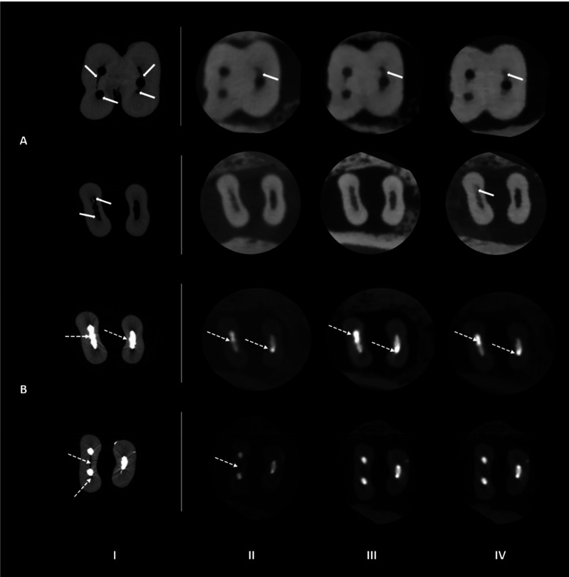
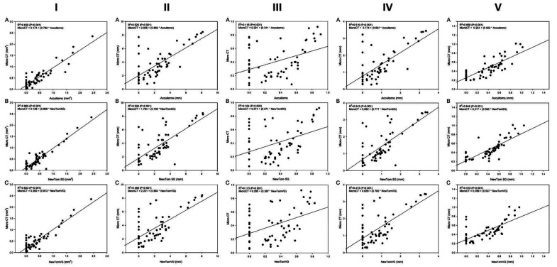
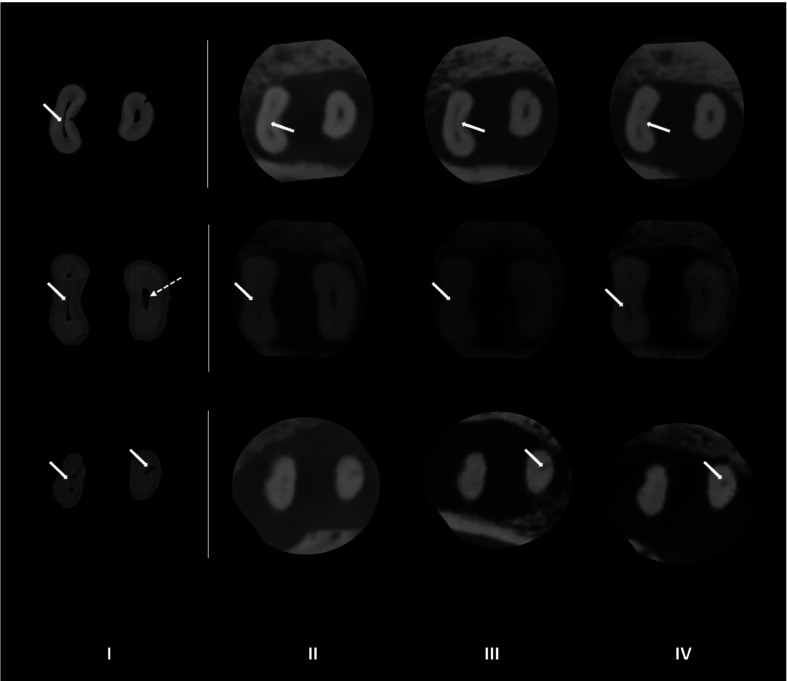
Materials and methods: Isthmus-containing mesial roots of mandibular molars (n=14) were scanned by micro-CT and 3 CBCT devices: 3D Accuitomo 170 (ACC), NewTom 5G (N5G) and NewTom VGi evo (NEVO). Two calibrated observers evaluated the images for 2-dimensional quantitative parameters, the presence of debris or root perforation, and filling quality in the root canal and isthmus. The kappa coefficient, analysis of variance, and the Tukey test were used for statistical analyses (α=5%).
Results: Substantial intra-observer agreement (κ=0.63) was found between micro-CT and ACC, N5G, and NEVO. Debris detection was difficult using ACC (42.9%), N5G (40.0%), and NEVO (40%), with no agreement between micro-CT and ACC, N5G, and NEVO (0.05<κ<0.12). After biomechanical preparation, 2.4%-4.8% of CBCT images showed root perforation that was absent on micro-CT. The 2D parameters showed satisfactory reproducibility between micro-CT and ACC, N5G, and NEVO (intraclass correlation coefficient: 0.60-0.73). Partially filled isthmuses were observed in 2.9% of the ACC images, 8.8% of the N5G and NEVO images, and 26.5% of the micro-CT images, with no agreement between micro-CT and ACC, and poor agreement between micro-CT and N5G and NEVO. Excellent agreement was found for area, perimeter, and the major and minor diameters, while the roundness measures were satisfactory.
Conclusion: CBCT images aided in isthmus detection and classification, but did not allow their classification after biomechanical preparation and root canal filling.


 分享
分享
 求助内容:
求助内容: 应助结果提醒方式:
 扫码关注我们
