Han-Gyeol Yeom, Wan Lee, Su-Il Han, Jae-Hoon Lee, Byung-Do Lee
{"title":"Mucocele in the maxillary sinus involving the orbit: A report of 2 cases.","authors":"Han-Gyeol Yeom, Wan Lee, Su-Il Han, Jae-Hoon Lee, Byung-Do Lee","doi":"10.5624/isd.20210278","DOIUrl":null,"url":null,"abstract":"<p><p>Mucocele of the paranasal sinuses is a benign, slow-growing, expansile lesion. Maxillary sinus mucoceles are usually associated with painless bulging of the cheek; however, orbital expansion is rarely observed. Maxillary sinus mucoceles can be classified as primary or secondary according to their etiology. An impediment to sinus ostium ventilation is thought to be the cause of primary mucocele, while sequestering of residual mucosa after surgery in the wound and long-term retention of tissue fluid have been suggested to lead to the formation of secondary mucocele. This report presents 2 cases of primary and secondary mucoceles, with a focus on radiographic features. As primary and superiorly positioned secondary maxillary sinus mucoceles are uncommon and their close proximity to the orbit predisposes the patient to significant morbidity, the authors expect that this report will contribute to a better understanding and diagnosis of maxillary sinus mucocele involving the orbit.</p>","PeriodicalId":51714,"journal":{"name":"Imaging Science in Dentistry","volume":"52 3","pages":"327-332"},"PeriodicalIF":2.1000,"publicationDate":"2022-09-01","publicationTypes":"Journal Article","fieldsOfStudy":null,"isOpenAccess":false,"openAccessPdf":"https://ftp.ncbi.nlm.nih.gov/pub/pmc/oa_pdf/9d/29/isd-52-327.PMC9530297.pdf","citationCount":"1","resultStr":null,"platform":"Semanticscholar","paperid":null,"PeriodicalName":"Imaging Science in Dentistry","FirstCategoryId":"1085","ListUrlMain":"https://doi.org/10.5624/isd.20210278","RegionNum":0,"RegionCategory":null,"ArticlePicture":[],"TitleCN":null,"AbstractTextCN":null,"PMCID":null,"EPubDate":"2022/6/2 0:00:00","PubModel":"Epub","JCR":"Q3","JCRName":"DENTISTRY, ORAL SURGERY & MEDICINE","Score":null,"Total":0}
引用次数: 1
Abstract
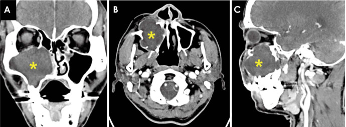
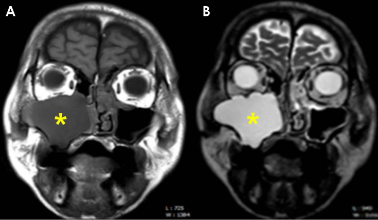
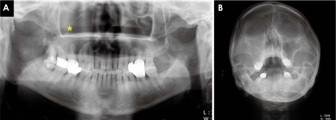
Mucocele of the paranasal sinuses is a benign, slow-growing, expansile lesion. Maxillary sinus mucoceles are usually associated with painless bulging of the cheek; however, orbital expansion is rarely observed. Maxillary sinus mucoceles can be classified as primary or secondary according to their etiology. An impediment to sinus ostium ventilation is thought to be the cause of primary mucocele, while sequestering of residual mucosa after surgery in the wound and long-term retention of tissue fluid have been suggested to lead to the formation of secondary mucocele. This report presents 2 cases of primary and secondary mucoceles, with a focus on radiographic features. As primary and superiorly positioned secondary maxillary sinus mucoceles are uncommon and their close proximity to the orbit predisposes the patient to significant morbidity, the authors expect that this report will contribute to a better understanding and diagnosis of maxillary sinus mucocele involving the orbit.


 分享
分享
 求助内容:
求助内容: 应助结果提醒方式:
 扫码关注我们
