{"title":"Scalloped border as a possible diagnostic aid for differentiating jaw lesions: A pictorial essay.","authors":"Hamed Mortazavi, Maryam Baharvand, Yaser Safi","doi":"10.5624/isd.20220033","DOIUrl":null,"url":null,"abstract":"<p><strong>Purpose: </strong>The aim of this study was to introduce a category of jaw lesions comprising cysts and tumors associated with scalloped borders.</p><p><strong>Materials and methods: </strong>General search engines and specialized databases including Google Scholar, PubMed, PubMed Central, and Scopus, as well as an authoritative textbook, were used to find relevant studies by using keywords such as \"jaw lesion,\" \"jaw disease,\" \"scalloping,\" \"scalloped border,\" \"scalloped margin,\" \"irregular border,\" and \"irregular margin.\" Out of 289 articles, 252 records were removed because they were duplicates, did not have a relevant title, or did not mention the frequency of findings described using the term \"scalloped border.\" Finally, 37 closely related articles were chosen.</p><p><strong>Results: </strong>According to the relevant literature, scalloped borders are found most frequently in ameloblastoma, followed by simple bone cyst, central giant cell granuloma, odontogenic keratocyst, and glandular odontogenic cyst.</p><p><strong>Conclusion: </strong>The lesions most frequently reported to have scalloped borders are ameloblastoma, central giant cell granuloma, odontogenic keratocyst, simple bone cyst, and glandular odontogenic cyst.</p>","PeriodicalId":51714,"journal":{"name":"Imaging Science in Dentistry","volume":"52 3","pages":"309-317"},"PeriodicalIF":2.1000,"publicationDate":"2022-09-01","publicationTypes":"Journal Article","fieldsOfStudy":null,"isOpenAccess":false,"openAccessPdf":"https://ftp.ncbi.nlm.nih.gov/pub/pmc/oa_pdf/a9/0b/isd-52-309.PMC9530295.pdf","citationCount":"0","resultStr":null,"platform":"Semanticscholar","paperid":null,"PeriodicalName":"Imaging Science in Dentistry","FirstCategoryId":"1085","ListUrlMain":"https://doi.org/10.5624/isd.20220033","RegionNum":0,"RegionCategory":null,"ArticlePicture":[],"TitleCN":null,"AbstractTextCN":null,"PMCID":null,"EPubDate":"2022/7/5 0:00:00","PubModel":"Epub","JCR":"Q3","JCRName":"DENTISTRY, ORAL SURGERY & MEDICINE","Score":null,"Total":0}
引用次数: 0
Abstract
Purpose: The aim of this study was to introduce a category of jaw lesions comprising cysts and tumors associated with scalloped borders.
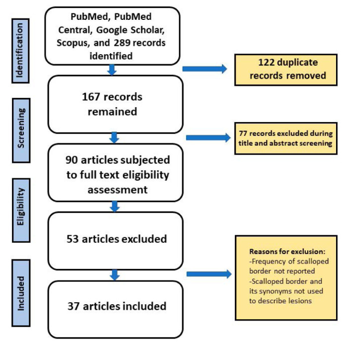
Materials and methods: General search engines and specialized databases including Google Scholar, PubMed, PubMed Central, and Scopus, as well as an authoritative textbook, were used to find relevant studies by using keywords such as "jaw lesion," "jaw disease," "scalloping," "scalloped border," "scalloped margin," "irregular border," and "irregular margin." Out of 289 articles, 252 records were removed because they were duplicates, did not have a relevant title, or did not mention the frequency of findings described using the term "scalloped border." Finally, 37 closely related articles were chosen.
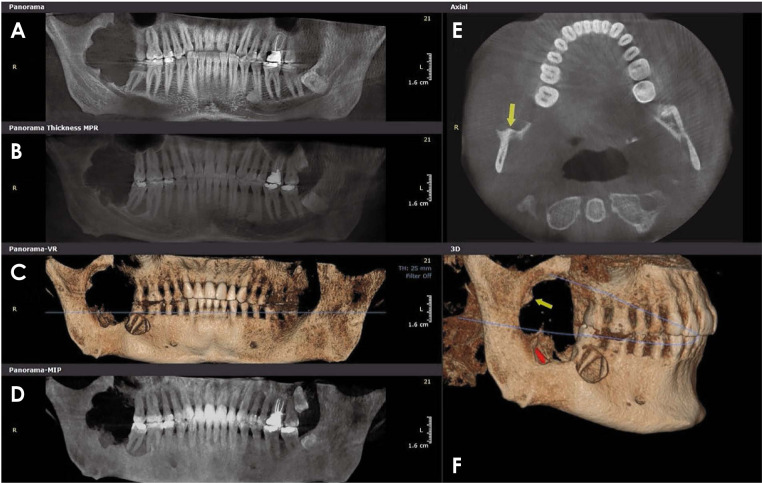
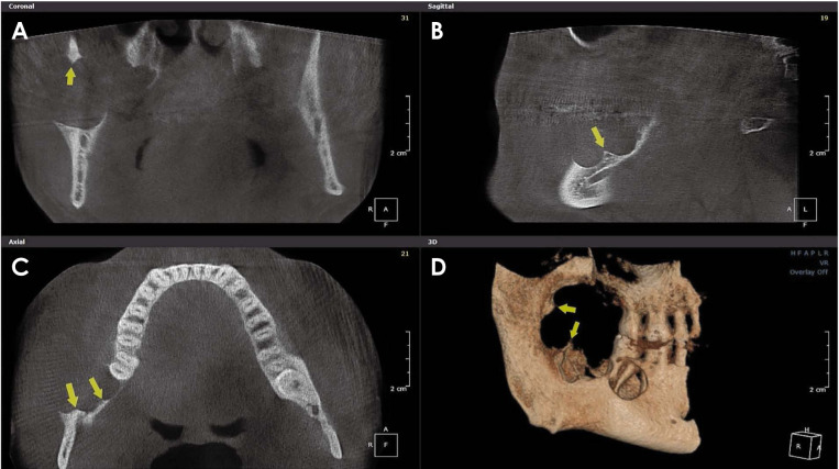
Results: According to the relevant literature, scalloped borders are found most frequently in ameloblastoma, followed by simple bone cyst, central giant cell granuloma, odontogenic keratocyst, and glandular odontogenic cyst.
Conclusion: The lesions most frequently reported to have scalloped borders are ameloblastoma, central giant cell granuloma, odontogenic keratocyst, simple bone cyst, and glandular odontogenic cyst.


 分享
分享
 求助内容:
求助内容: 应助结果提醒方式:
 扫码关注我们
