Influence of kilovoltage-peak and the metal artifact reduction tool in cone-beam computed tomography on the detection of bone defects around titanium-zirconia and zirconia implants.
Rocharles Cavalcante Fontenele, Eduarda Helena Leandro Nascimento, Ana Catarina Imbelloni-Vasconcelos, Luciano Augusto Cano Martins, Andrea Dos Anjos Pontual, Flávia Maria Moraes Ramos-Perez, Deborah Queiroz Freitas
{"title":"Influence of kilovoltage-peak and the metal artifact reduction tool in cone-beam computed tomography on the detection of bone defects around titanium-zirconia and zirconia implants.","authors":"Rocharles Cavalcante Fontenele, Eduarda Helena Leandro Nascimento, Ana Catarina Imbelloni-Vasconcelos, Luciano Augusto Cano Martins, Andrea Dos Anjos Pontual, Flávia Maria Moraes Ramos-Perez, Deborah Queiroz Freitas","doi":"10.5624/isd.20220040","DOIUrl":null,"url":null,"abstract":"<p><strong>Purpose: </strong>The aim of this study was to assess the influence of kilovoltage-peak (kVp) and the metal artifact reduction (MAR) tool on the detection of buccal and lingual peri-implant dehiscence in the presence of titanium-zirconia (Ti-Zr) and zirconia (Zr) implants in cone-beam computed tomography (CBCT) images.</p><p><strong>Materials and methods: </strong>Twenty implant sites were created in the posterior region of human mandibles, including control sites (without dehiscence) and experimental sites (with dehiscence). Individually, a Ti-Zr or Zr implant was placed in each implant site. CBCT scans were performed using a Picasso Trio device, with variation in the kVp setting (70 or 90 kVp) and whether the MAR tool was used. Three oral radiologists scored the detection of dehiscence using a 5-point scale. The area under the receiver operating characteristic (ROC) curve, sensitivity, and specificity were calculated and compared by multi-way analysis of variance (α=0.05).</p><p><strong>Results: </strong>The kVp, cortical plate involved (buccal or lingual cortices), and MAR did not influence any diagnostic values (<i>P</i>>0.05). The material of the implant did not influence the ROC curve values (<i>P</i>>0.05). In contrast, the sensitivity and specificity were statistically significantly influenced by the implant material (<i>P</i><0.05) with Zr implants showing higher sensitivity values and lower specificity values than Ti-Zr implants.</p><p><strong>Conclusion: </strong>The detection of peri-implant dehiscence was not influenced by kVp, use of the MAR tool, or the cortical plate. Greater sensitivity and lower specificity were shown for the detection of peri-implant dehiscence in the presence of a Zr implant.</p>","PeriodicalId":51714,"journal":{"name":"Imaging Science in Dentistry","volume":"52 3","pages":"267-273"},"PeriodicalIF":2.1000,"publicationDate":"2022-09-01","publicationTypes":"Journal Article","fieldsOfStudy":null,"isOpenAccess":false,"openAccessPdf":"https://ftp.ncbi.nlm.nih.gov/pub/pmc/oa_pdf/50/50/isd-52-267.PMC9530290.pdf","citationCount":"1","resultStr":null,"platform":"Semanticscholar","paperid":null,"PeriodicalName":"Imaging Science in Dentistry","FirstCategoryId":"1085","ListUrlMain":"https://doi.org/10.5624/isd.20220040","RegionNum":0,"RegionCategory":null,"ArticlePicture":[],"TitleCN":null,"AbstractTextCN":null,"PMCID":null,"EPubDate":"2022/7/5 0:00:00","PubModel":"Epub","JCR":"Q3","JCRName":"DENTISTRY, ORAL SURGERY & MEDICINE","Score":null,"Total":0}
引用次数: 1
Abstract
Purpose: The aim of this study was to assess the influence of kilovoltage-peak (kVp) and the metal artifact reduction (MAR) tool on the detection of buccal and lingual peri-implant dehiscence in the presence of titanium-zirconia (Ti-Zr) and zirconia (Zr) implants in cone-beam computed tomography (CBCT) images.
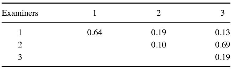
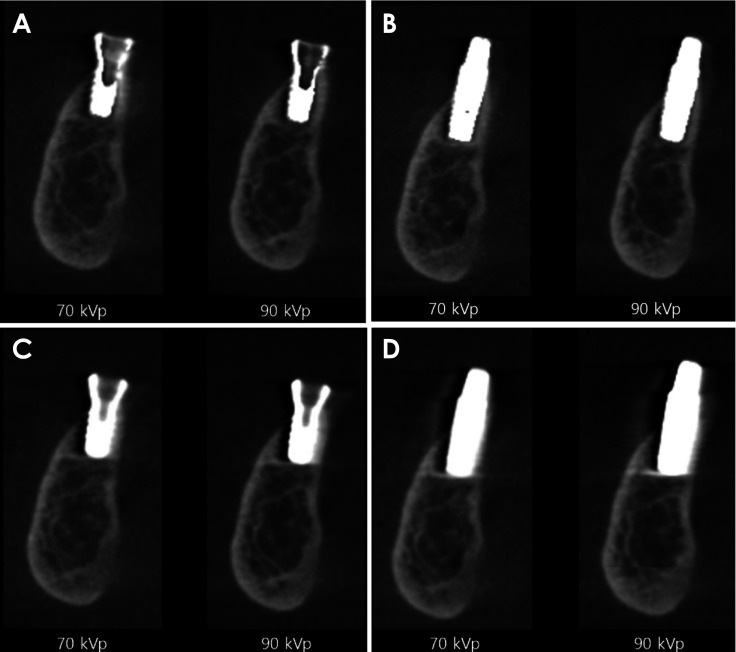
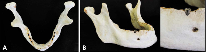
Materials and methods: Twenty implant sites were created in the posterior region of human mandibles, including control sites (without dehiscence) and experimental sites (with dehiscence). Individually, a Ti-Zr or Zr implant was placed in each implant site. CBCT scans were performed using a Picasso Trio device, with variation in the kVp setting (70 or 90 kVp) and whether the MAR tool was used. Three oral radiologists scored the detection of dehiscence using a 5-point scale. The area under the receiver operating characteristic (ROC) curve, sensitivity, and specificity were calculated and compared by multi-way analysis of variance (α=0.05).
Results: The kVp, cortical plate involved (buccal or lingual cortices), and MAR did not influence any diagnostic values (P>0.05). The material of the implant did not influence the ROC curve values (P>0.05). In contrast, the sensitivity and specificity were statistically significantly influenced by the implant material (P<0.05) with Zr implants showing higher sensitivity values and lower specificity values than Ti-Zr implants.
Conclusion: The detection of peri-implant dehiscence was not influenced by kVp, use of the MAR tool, or the cortical plate. Greater sensitivity and lower specificity were shown for the detection of peri-implant dehiscence in the presence of a Zr implant.


 分享
分享
 求助内容:
求助内容: 应助结果提醒方式:
 扫码关注我们
