Dae-Gwang Yoo, Shin Hwang, Dae-Wook Hwang, Ki-Hun Kim, Chul-Soo Ahn, Tae-Yong Ha, Gi-Won Song, Dong-Hwan Jung, Gil-Chun Park, Sung-Gyu Lee
{"title":"Case report of a pancreatic squamoid cyst.","authors":"Dae-Gwang Yoo, Shin Hwang, Dae-Wook Hwang, Ki-Hun Kim, Chul-Soo Ahn, Tae-Yong Ha, Gi-Won Song, Dong-Hwan Jung, Gil-Chun Park, Sung-Gyu Lee","doi":"10.14701/kjhbps.2013.17.4.181","DOIUrl":null,"url":null,"abstract":"<p><p>Squamoid cyst of the pancreas is a very rare disease and it has been proposed only recently as a distinct pathologic lesion. We herein present a case of pancreatic squamoid cyst in a patient who underwent laparoscopic resection. A 60-year-old woman had an abdominal computed tomography (CT) scan for a routine check-up, and a multi-cystic lesion of 1.8-cm in size was incidentally found in the tail of the pancreas. Biochemical laboratory tests were within normal limits. At first, we presumed that the most likely diagnosis of the cystic lesion was an intraductal papillary mucinous neoplasm. To treat this lesion, we performed laparoscopic spleen-saving distal pancreatectomy. The patient showed the usual routine postoperative course and she was discharged 10 days after surgery. On examination of the resected specimen, a well-defined, oligolocular cystic mass was found in the pancreatic tail, without a solid portion. Histologic examination revealed that the cysts had linings ranging from flat squamoid cells to transitional cells with non-keratinization. After immunohistochemical staining, the final diagnosis was confirmed to be squamoid cyst of the pancreas. This lesion appears to be regarded as a benign entity, thus an extended operation should be avoided and resection of the lesion can be performed minimally. </p>","PeriodicalId":91136,"journal":{"name":"Korean journal of hepato-biliary-pancreatic surgery","volume":"17 4","pages":"181-5"},"PeriodicalIF":0.0000,"publicationDate":"2013-11-01","publicationTypes":"Journal Article","fieldsOfStudy":null,"isOpenAccess":false,"openAccessPdf":"https://sci-hub-pdf.com/10.14701/kjhbps.2013.17.4.181","citationCount":"10","resultStr":null,"platform":"Semanticscholar","paperid":null,"PeriodicalName":"Korean journal of hepato-biliary-pancreatic surgery","FirstCategoryId":"1085","ListUrlMain":"https://doi.org/10.14701/kjhbps.2013.17.4.181","RegionNum":0,"RegionCategory":null,"ArticlePicture":[],"TitleCN":null,"AbstractTextCN":null,"PMCID":null,"EPubDate":"2013/11/20 0:00:00","PubModel":"Epub","JCR":"","JCRName":"","Score":null,"Total":0}
引用次数: 10
Abstract
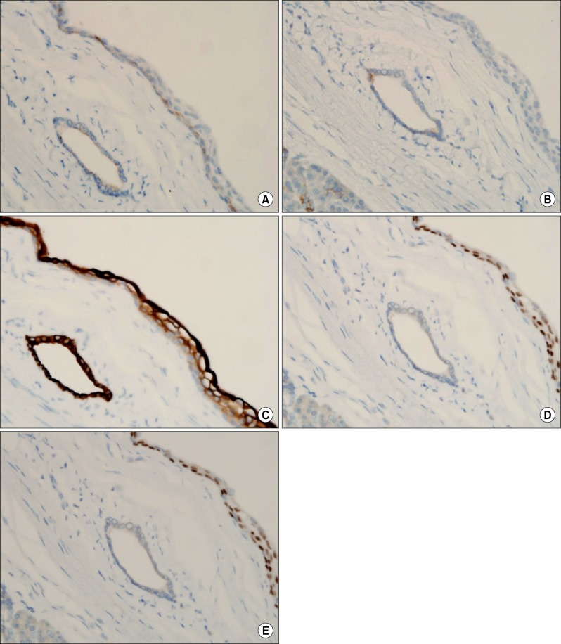
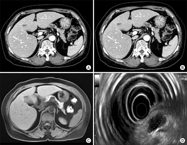
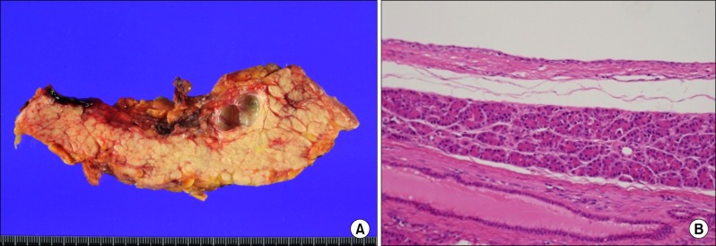
Squamoid cyst of the pancreas is a very rare disease and it has been proposed only recently as a distinct pathologic lesion. We herein present a case of pancreatic squamoid cyst in a patient who underwent laparoscopic resection. A 60-year-old woman had an abdominal computed tomography (CT) scan for a routine check-up, and a multi-cystic lesion of 1.8-cm in size was incidentally found in the tail of the pancreas. Biochemical laboratory tests were within normal limits. At first, we presumed that the most likely diagnosis of the cystic lesion was an intraductal papillary mucinous neoplasm. To treat this lesion, we performed laparoscopic spleen-saving distal pancreatectomy. The patient showed the usual routine postoperative course and she was discharged 10 days after surgery. On examination of the resected specimen, a well-defined, oligolocular cystic mass was found in the pancreatic tail, without a solid portion. Histologic examination revealed that the cysts had linings ranging from flat squamoid cells to transitional cells with non-keratinization. After immunohistochemical staining, the final diagnosis was confirmed to be squamoid cyst of the pancreas. This lesion appears to be regarded as a benign entity, thus an extended operation should be avoided and resection of the lesion can be performed minimally.


 分享
分享

 求助内容:
求助内容: 应助结果提醒方式:
 扫码关注我们
