Abdullateef A. Alzolibani , Zafar Rasheed , Ghada Bin Saif , Mohammed S. Al-Dhubaibi , Ahmad A. Al Robaee
{"title":"Altered expression of intracellular Toll-like receptors in peripheral blood mononuclear cells from patients with alopecia areata","authors":"Abdullateef A. Alzolibani , Zafar Rasheed , Ghada Bin Saif , Mohammed S. Al-Dhubaibi , Ahmad A. Al Robaee","doi":"10.1016/j.bbacli.2016.03.006","DOIUrl":null,"url":null,"abstract":"<div><h3>Background</h3><p>Toll-like receptors (TLRs) are pattern-recognition-receptors that sense a variety of pathogens and initiation of innate and adaptive immune responses. This study was undertaken to investigate the expression of TLRs in peripheral blood-mononuclear cells (PBMCs) of AA patients and to determine whether TLR-mediated inflammatory signals are important for the perspective of AA management.</p></div><div><h3>Methods</h3><p>Gene expression of TLRs and T-helper (Th) type-1, Th-2, Th-17 and regulatory T-cell cytokines in PBMCs was quantified by TaqMan Assays. Production of these cytokines in serum samples was determined by sandwich ELISAs.</p></div><div><h3>Results</h3><p>All TLRs (TLRs 1–10) were expressed in PBMCs of AA patients. Importantly intracellular TLRs (TLRs 3, 7, 8 and 9) were significantly up-regulated in AA patients as compared with controls (p<!--> <!--><<!--> <!-->0.05). Interleukin (IL)-2, TNF-α, and IL-17A gene expression in patients' PBMCs and their secretion in patients' sera were significantly higher as compared with their respective controls (p<!--> <!--><<!--> <!-->0.05). Whereas, TGF-β gene expression in patients' PBMCs and TGF-β protein level in patients' sera were significantly lower as compared with their controls (p<!--> <!--><<!--> <!-->0.05).</p></div><div><h3>Conclusion</h3><p>This is the first report that shows the comprehensive expression profile of TLRs in AA patients. We conclude that up-regulated expression of intracellular TLRs in PBMCs of AA patients may play an active role in abnormal regulation of Th-1, Th-17 and regulatory T-cell cytokines in alopecia areata.</p></div><div><h3>General significance</h3><p>Targeting of TLRs and their associated inflammatory signaling will open new areas of research; this may lead to the development of novel therapeutic targets for the treatment of AA or other skin disorders.</p></div>","PeriodicalId":72344,"journal":{"name":"BBA clinical","volume":"5 ","pages":"Pages 134-142"},"PeriodicalIF":0.0000,"publicationDate":"2016-06-01","publicationTypes":"Journal Article","fieldsOfStudy":null,"isOpenAccess":false,"openAccessPdf":"https://sci-hub-pdf.com/10.1016/j.bbacli.2016.03.006","citationCount":"13","resultStr":null,"platform":"Semanticscholar","paperid":null,"PeriodicalName":"BBA clinical","FirstCategoryId":"1085","ListUrlMain":"https://www.sciencedirect.com/science/article/pii/S2214647416300101","RegionNum":0,"RegionCategory":null,"ArticlePicture":[],"TitleCN":null,"AbstractTextCN":null,"PMCID":null,"EPubDate":"","PubModel":"","JCR":"","JCRName":"","Score":null,"Total":0}
引用次数: 13
Abstract
Background
Toll-like receptors (TLRs) are pattern-recognition-receptors that sense a variety of pathogens and initiation of innate and adaptive immune responses. This study was undertaken to investigate the expression of TLRs in peripheral blood-mononuclear cells (PBMCs) of AA patients and to determine whether TLR-mediated inflammatory signals are important for the perspective of AA management.
Methods
Gene expression of TLRs and T-helper (Th) type-1, Th-2, Th-17 and regulatory T-cell cytokines in PBMCs was quantified by TaqMan Assays. Production of these cytokines in serum samples was determined by sandwich ELISAs.
Results
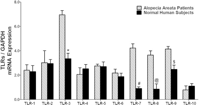
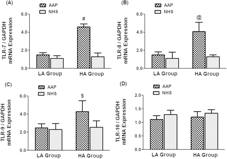
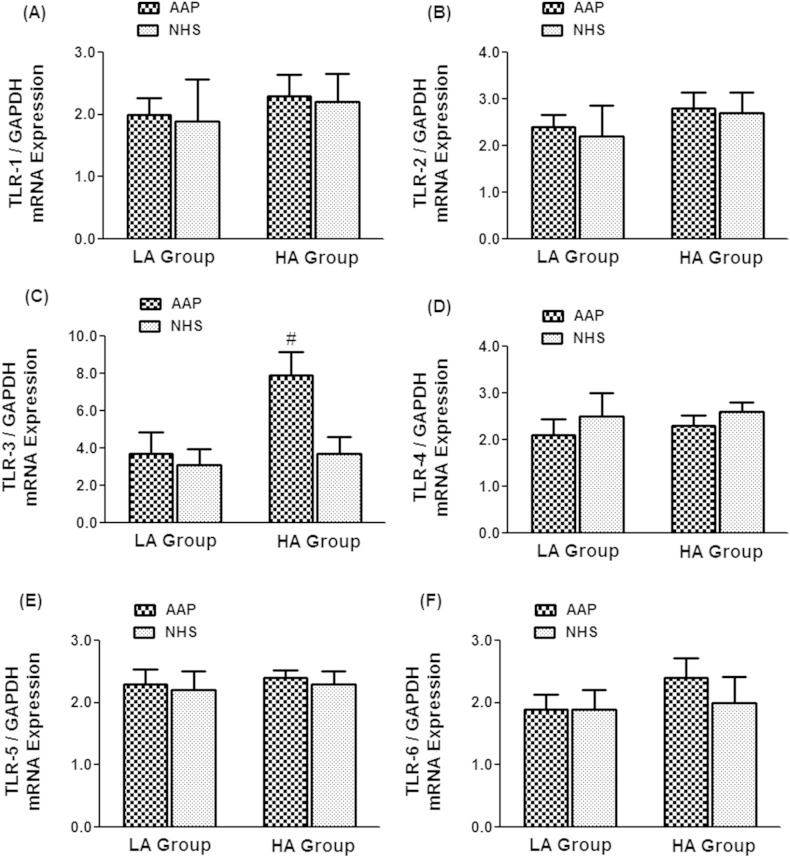
All TLRs (TLRs 1–10) were expressed in PBMCs of AA patients. Importantly intracellular TLRs (TLRs 3, 7, 8 and 9) were significantly up-regulated in AA patients as compared with controls (p < 0.05). Interleukin (IL)-2, TNF-α, and IL-17A gene expression in patients' PBMCs and their secretion in patients' sera were significantly higher as compared with their respective controls (p < 0.05). Whereas, TGF-β gene expression in patients' PBMCs and TGF-β protein level in patients' sera were significantly lower as compared with their controls (p < 0.05).
Conclusion
This is the first report that shows the comprehensive expression profile of TLRs in AA patients. We conclude that up-regulated expression of intracellular TLRs in PBMCs of AA patients may play an active role in abnormal regulation of Th-1, Th-17 and regulatory T-cell cytokines in alopecia areata.
General significance
Targeting of TLRs and their associated inflammatory signaling will open new areas of research; this may lead to the development of novel therapeutic targets for the treatment of AA or other skin disorders.


 分享
分享

 求助内容:
求助内容: 应助结果提醒方式:
 扫码关注我们
