{"title":"Glycosaminoglycan measured from synovial fluid serves as a useful indicator for progression of Osteoarthritis and complements Kellgren–Lawrence Score","authors":"Priya Kulkarni , Shantanu Deshpande , Soumya Koppikar , Sanjay Patil , Dhanashri Ingale , Abhay Harsulkar","doi":"10.1016/j.bbacli.2016.05.002","DOIUrl":null,"url":null,"abstract":"<div><h3>Background</h3><p>Plain radiography is the first choice for diagnosis and monitoring of knee-osteoarthritis (OA) while, Kellgren–Lawrence score (KL) is most widely used to grade OA severity. However, incompetency for reproducibility of joint space measurement in longitudinal assessment and non-linearity of KL-score system, limits radiography-based early diagnosis of the disease. Glycosaminoglycan (GAG) is direct cartilage-degradation product, which can be measured biochemically. We strived to correlate KL-score and GAG from OA patients to compliment KL-system.</p></div><div><h3>Methods</h3><p>We obtained 34 synovial-fluid (SF) samples from 28 OA patients (few bilateral) with different disease severity using arthrocetesis. All patients were categorised using radiographic KL-score-system. SFs were further analysed for GAG estimation using 1,2-dimethylmethylene blue (DMMB) assay.</p></div><div><h3>Results</h3><p>A substantial increase in GAG was noted in KL-grade-II and III, comparing grade-I patients, indicating amplified cartilage-degradation. KL-grade-IV patients revealed further rise in GAG reflecting more cartilage-loss. Another category of grade-IV patients with lower GAG were also detected, indicating close to total cartilage-loss.</p></div><div><h3>Conclusions</h3><p>Accurate diagnosis of cartilage-loss remains a challenge with OA due to limitations of KL-system; thus no target intervention is available to arrest active cartilage-loss. We propose, GAG-estimation in OA patients, characterizes accurate biochemical depiction of cartilage degeneration. General Significance: Radiology often fails to reveal an accurate cartilage loss, associated with OA. GAG levels from the SFs of OA patients' serve as a useful marker, which parallels cartilage degeneration and strengthen radiographic grading system, ultimately</p></div>","PeriodicalId":72344,"journal":{"name":"BBA clinical","volume":"6 ","pages":"Pages 1-4"},"PeriodicalIF":0.0000,"publicationDate":"2016-12-01","publicationTypes":"Journal Article","fieldsOfStudy":null,"isOpenAccess":false,"openAccessPdf":"https://sci-hub-pdf.com/10.1016/j.bbacli.2016.05.002","citationCount":"19","resultStr":null,"platform":"Semanticscholar","paperid":null,"PeriodicalName":"BBA clinical","FirstCategoryId":"1085","ListUrlMain":"https://www.sciencedirect.com/science/article/pii/S221464741630023X","RegionNum":0,"RegionCategory":null,"ArticlePicture":[],"TitleCN":null,"AbstractTextCN":null,"PMCID":null,"EPubDate":"","PubModel":"","JCR":"","JCRName":"","Score":null,"Total":0}
引用次数: 19
Abstract
Background
Plain radiography is the first choice for diagnosis and monitoring of knee-osteoarthritis (OA) while, Kellgren–Lawrence score (KL) is most widely used to grade OA severity. However, incompetency for reproducibility of joint space measurement in longitudinal assessment and non-linearity of KL-score system, limits radiography-based early diagnosis of the disease. Glycosaminoglycan (GAG) is direct cartilage-degradation product, which can be measured biochemically. We strived to correlate KL-score and GAG from OA patients to compliment KL-system.
Methods
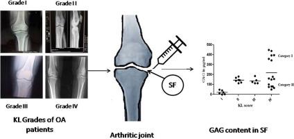
We obtained 34 synovial-fluid (SF) samples from 28 OA patients (few bilateral) with different disease severity using arthrocetesis. All patients were categorised using radiographic KL-score-system. SFs were further analysed for GAG estimation using 1,2-dimethylmethylene blue (DMMB) assay.
Results
A substantial increase in GAG was noted in KL-grade-II and III, comparing grade-I patients, indicating amplified cartilage-degradation. KL-grade-IV patients revealed further rise in GAG reflecting more cartilage-loss. Another category of grade-IV patients with lower GAG were also detected, indicating close to total cartilage-loss.
Conclusions
Accurate diagnosis of cartilage-loss remains a challenge with OA due to limitations of KL-system; thus no target intervention is available to arrest active cartilage-loss. We propose, GAG-estimation in OA patients, characterizes accurate biochemical depiction of cartilage degeneration. General Significance: Radiology often fails to reveal an accurate cartilage loss, associated with OA. GAG levels from the SFs of OA patients' serve as a useful marker, which parallels cartilage degeneration and strengthen radiographic grading system, ultimately


 分享
分享

 求助内容:
求助内容: 应助结果提醒方式:
 扫码关注我们
