Richard Laforest, Farrokh Dehdashti, Yongjian Liu, Jennifer Frye, Sarah Frye, Hannah Luehmann, Deborah Sultan, Joseph S Shan, Bruce D Freimark, Barry A Siegel
{"title":"First-in-Man Evaluation of <sup>124</sup>I-PGN650: A PET Tracer for Detecting Phosphatidylserine as a Biomarker of the Solid Tumor Microenvironment.","authors":"Richard Laforest, Farrokh Dehdashti, Yongjian Liu, Jennifer Frye, Sarah Frye, Hannah Luehmann, Deborah Sultan, Joseph S Shan, Bruce D Freimark, Barry A Siegel","doi":"10.1177/1536012117733349","DOIUrl":null,"url":null,"abstract":"<p><strong>Purpose: </strong>PGN650 is a F(ab')<sub>2</sub> antibody fragment that targets phosphatidylserine (PS), a marker normally absent that becomes exposed on tumor cells and tumor vasculature in response to oxidative stress and increases in response to therapy. PGN650 was labeled with <sup>124</sup>I to create a positron emission tomography (PET) agent as an in vivo biomarker for tumor microenvironment and response to therapy. In this phase 0 study, we evaluated the pharmacokinetics, safety, radiation dosimetry, and tumor targeting of this tracer in a cohort of patients with cancer.</p><p><strong>Methods: </strong>Eleven patients with known solid tumors received approximately 140 MBq (3.8 mCi) <sup>124</sup>I-PGN650 intravenously and underwent positron emission tomography-computed tomography (PET/CT) approximately 1 hour, 3 hours, and either 24 hours or 48 hours later to establish tracer kinetics for the purpose of calculating radiation dosimetry (from integration of the organ time-activity curves and OLINDA/EXM using the adult male and female models).</p><p><strong>Results: </strong>Known tumor foci demonstrated mildly increased uptake, with the highest activity at the latest imaging time. There were no unexpected adverse events. The liver was the organ receiving the highest radiation dose (0.77 mGy/MBq); the effective dose was 0.41 mSv/MBq.</p><p><strong>Conclusion: </strong>Although <sup>124</sup>I-PGN650 is safe for human PET imaging, the tumor targeting with this agent in patients was less than previously observed in animal studies.</p>","PeriodicalId":18855,"journal":{"name":"Molecular Imaging","volume":"16 ","pages":"1536012117733349"},"PeriodicalIF":2.4000,"publicationDate":"2017-01-01","publicationTypes":"Journal Article","fieldsOfStudy":null,"isOpenAccess":false,"openAccessPdf":"https://ftp.ncbi.nlm.nih.gov/pub/pmc/oa_pdf/c4/e8/10.1177_1536012117733349.PMC5648081.pdf","citationCount":"0","resultStr":null,"platform":"Semanticscholar","paperid":null,"PeriodicalName":"Molecular Imaging","FirstCategoryId":"3","ListUrlMain":"https://doi.org/10.1177/1536012117733349","RegionNum":4,"RegionCategory":"医学","ArticlePicture":[],"TitleCN":null,"AbstractTextCN":null,"PMCID":null,"EPubDate":"","PubModel":"","JCR":"Q3","JCRName":"BIOCHEMICAL RESEARCH METHODS","Score":null,"Total":0}
引用次数: 0
Abstract
Purpose: PGN650 is a F(ab')2 antibody fragment that targets phosphatidylserine (PS), a marker normally absent that becomes exposed on tumor cells and tumor vasculature in response to oxidative stress and increases in response to therapy. PGN650 was labeled with 124I to create a positron emission tomography (PET) agent as an in vivo biomarker for tumor microenvironment and response to therapy. In this phase 0 study, we evaluated the pharmacokinetics, safety, radiation dosimetry, and tumor targeting of this tracer in a cohort of patients with cancer.
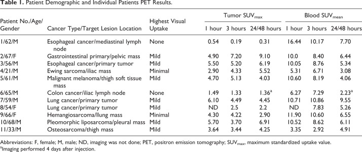
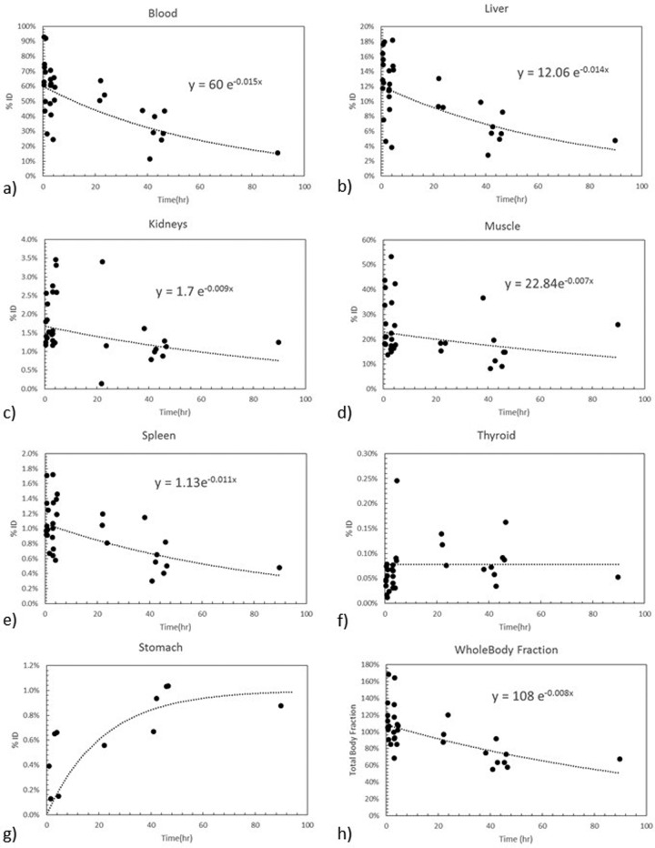
Methods: Eleven patients with known solid tumors received approximately 140 MBq (3.8 mCi) 124I-PGN650 intravenously and underwent positron emission tomography-computed tomography (PET/CT) approximately 1 hour, 3 hours, and either 24 hours or 48 hours later to establish tracer kinetics for the purpose of calculating radiation dosimetry (from integration of the organ time-activity curves and OLINDA/EXM using the adult male and female models).
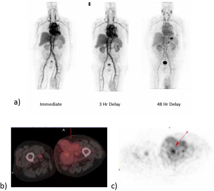
Results: Known tumor foci demonstrated mildly increased uptake, with the highest activity at the latest imaging time. There were no unexpected adverse events. The liver was the organ receiving the highest radiation dose (0.77 mGy/MBq); the effective dose was 0.41 mSv/MBq.
Conclusion: Although 124I-PGN650 is safe for human PET imaging, the tumor targeting with this agent in patients was less than previously observed in animal studies.
Molecular ImagingBiochemistry, Genetics and Molecular Biology-Biotechnology
自引率
3.60%
发文量
21
期刊介绍:
Molecular Imaging is a peer-reviewed, open access journal highlighting the breadth of molecular imaging research from basic science to preclinical studies to human applications. This serves both the scientific and clinical communities by disseminating novel results and concepts relevant to the biological study of normal and disease processes in both basic and translational studies ranging from mice to humans.


 分享
分享
 求助内容:
求助内容: 应助结果提醒方式:
 扫码关注我们
