Evaluation of the Effect of Buccolingual and Apicocoronal Positions of Dental Implants on Stress and Strain in Alveolar Bone by Finite Element Analysis.
Farhood Massoumi, Mina Taheri, Abolghasem Mohammadi, Omid Amelirad
{"title":"Evaluation of the Effect of Buccolingual and Apicocoronal Positions of Dental Implants on Stress and Strain in Alveolar Bone by Finite Element Analysis.","authors":"Farhood Massoumi, Mina Taheri, Abolghasem Mohammadi, Omid Amelirad","doi":"","DOIUrl":null,"url":null,"abstract":"<p><strong>Objectives: </strong>The position of dental implants in the alveolar bone can affect the surrounding bone from biomechanical and biological aspects. The purpose of this study was to evaluate the effect of implant position on stress and strain distribution in the surrounding bone by using finite element analysis (FEA).</p><p><strong>Materials and methods: </strong>Thirteen computerized models of a 3.8-mm-diameter XiVE implant with the abutment and crown of a mandibular second premolar in a mandibular bone segment were designed. In the reference model, the implant was placed at the center of the alveolar ridge with its crest module located above the alveolar crest. In the other models, the implants were positioned buccally, lingually, coronally or apically by 0.5, 1 or 1.5mm. By using the ANSYS software program, a 100-N load was applied to the buccal cusp parallel to and at a 30-degree angle relative to the longitudinal axis of the fixture. The models were analyzed in terms of the distribution of stress and strain in the bone.</p><p><strong>Results: </strong>The different implant positions induced nonlinear stress and strain changes in the bone. The central, 1.5-mm apical, and 1.5-mm coronal implant positions induced high amounts of stress and strain under off-axial loads.</p><p><strong>Conclusions: </strong>Within the limitations of this study, the results showed that the stress and strain in the bone around the implant undergo small nonlinear changes with buccolingual and apicocoronal shifting of the implant and can be affected by the configuration of the implant in contact with the bone.</p>","PeriodicalId":30286,"journal":{"name":"Journal of Dentistry of Tehran University of Medical Sciences","volume":"15 1","pages":"10-19"},"PeriodicalIF":0.0000,"publicationDate":"2018-01-01","publicationTypes":"Journal Article","fieldsOfStudy":null,"isOpenAccess":false,"openAccessPdf":"https://www.ncbi.nlm.nih.gov/pmc/articles/PMC6026103/pdf/","citationCount":"0","resultStr":null,"platform":"Semanticscholar","paperid":null,"PeriodicalName":"Journal of Dentistry of Tehran University of Medical Sciences","FirstCategoryId":"1085","ListUrlMain":"","RegionNum":0,"RegionCategory":null,"ArticlePicture":[],"TitleCN":null,"AbstractTextCN":null,"PMCID":null,"EPubDate":"","PubModel":"","JCR":"","JCRName":"","Score":null,"Total":0}
引用次数: 0
Abstract
Objectives: The position of dental implants in the alveolar bone can affect the surrounding bone from biomechanical and biological aspects. The purpose of this study was to evaluate the effect of implant position on stress and strain distribution in the surrounding bone by using finite element analysis (FEA).
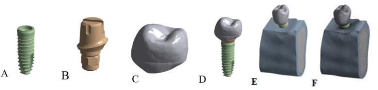
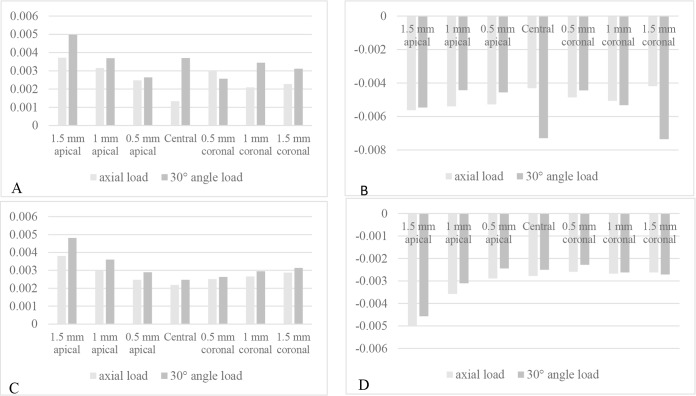
Materials and methods: Thirteen computerized models of a 3.8-mm-diameter XiVE implant with the abutment and crown of a mandibular second premolar in a mandibular bone segment were designed. In the reference model, the implant was placed at the center of the alveolar ridge with its crest module located above the alveolar crest. In the other models, the implants were positioned buccally, lingually, coronally or apically by 0.5, 1 or 1.5mm. By using the ANSYS software program, a 100-N load was applied to the buccal cusp parallel to and at a 30-degree angle relative to the longitudinal axis of the fixture. The models were analyzed in terms of the distribution of stress and strain in the bone.
Results: The different implant positions induced nonlinear stress and strain changes in the bone. The central, 1.5-mm apical, and 1.5-mm coronal implant positions induced high amounts of stress and strain under off-axial loads.
Conclusions: Within the limitations of this study, the results showed that the stress and strain in the bone around the implant undergo small nonlinear changes with buccolingual and apicocoronal shifting of the implant and can be affected by the configuration of the implant in contact with the bone.

 分享
分享


 求助内容:
求助内容: 应助结果提醒方式:
 扫码关注我们
