Examination of the vascularization of fetal kidney with three-dimensional power Doppler technique in pregnancies complicated by increased maternal blood pressure.
Andrea Suranyi, Miklos Nogrady, Abel Altorjay, Tibor Nyari, Gabor Nemeth
{"title":"Examination of the vascularization of fetal kidney with three-dimensional power Doppler technique in pregnancies complicated by increased maternal blood pressure.","authors":"Andrea Suranyi, Miklos Nogrady, Abel Altorjay, Tibor Nyari, Gabor Nemeth","doi":"10.1556/1646.10.2018.15","DOIUrl":null,"url":null,"abstract":"<p><p>The goal of this study was to investigate the fetal renal vascularization during the third trimester of gestation and the perinatal outcome in pregnancies diagnosed with hypertension. Depending on the medical history, the cases were divided into two groups: chronic hypertension (CHT) group and gestational hypertension (GHT) group. The vascularization and the volume of kidneys were observed in prenatal period by three-dimensional ultrasound. We monitored gestations and perinatal complications. Renal volume and vascularization were detected in 45 cases complicated by GHT and 21 cases with CHT during the 20-month study period. The alteration in fetal renal volume and vascularization may be an in utero cause of subsequent intrauterine and neonatal complications, such as cesarean section because of fetal distress (36%), perinatal infection (24%), treatment in neonatal intensive care unit (39%), or increased perinatal mortality (1%) in affected cases. The results demonstrate that fetuses with depressed vascularization of medullae had 1.5 times the risk of an abnormal outcome compared with the control group. The volume of kidneys had a strong correlation with their vascularization. Detailed ultrasound examinations of renal parenchyma appear to be useful for the prenatal diagnosis of intrauterine hypoxia, allowing the detection of potential pathological fetal conditions in utero.</p>","PeriodicalId":45181,"journal":{"name":"Interventional Medicine and Applied Science","volume":"10 1","pages":"7-12"},"PeriodicalIF":0.0000,"publicationDate":"2018-03-01","publicationTypes":"Journal Article","fieldsOfStudy":null,"isOpenAccess":false,"openAccessPdf":"https://sci-hub-pdf.com/10.1556/1646.10.2018.15","citationCount":"0","resultStr":null,"platform":"Semanticscholar","paperid":null,"PeriodicalName":"Interventional Medicine and Applied Science","FirstCategoryId":"1085","ListUrlMain":"https://doi.org/10.1556/1646.10.2018.15","RegionNum":0,"RegionCategory":null,"ArticlePicture":[],"TitleCN":null,"AbstractTextCN":null,"PMCID":null,"EPubDate":"","PubModel":"","JCR":"Q2","JCRName":"Medicine","Score":null,"Total":0}
引用次数: 0
Abstract
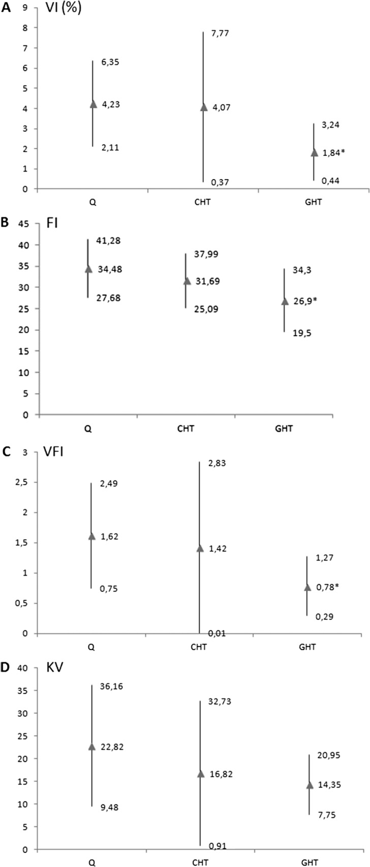
The goal of this study was to investigate the fetal renal vascularization during the third trimester of gestation and the perinatal outcome in pregnancies diagnosed with hypertension. Depending on the medical history, the cases were divided into two groups: chronic hypertension (CHT) group and gestational hypertension (GHT) group. The vascularization and the volume of kidneys were observed in prenatal period by three-dimensional ultrasound. We monitored gestations and perinatal complications. Renal volume and vascularization were detected in 45 cases complicated by GHT and 21 cases with CHT during the 20-month study period. The alteration in fetal renal volume and vascularization may be an in utero cause of subsequent intrauterine and neonatal complications, such as cesarean section because of fetal distress (36%), perinatal infection (24%), treatment in neonatal intensive care unit (39%), or increased perinatal mortality (1%) in affected cases. The results demonstrate that fetuses with depressed vascularization of medullae had 1.5 times the risk of an abnormal outcome compared with the control group. The volume of kidneys had a strong correlation with their vascularization. Detailed ultrasound examinations of renal parenchyma appear to be useful for the prenatal diagnosis of intrauterine hypoxia, allowing the detection of potential pathological fetal conditions in utero.


 分享
分享
 求助内容:
求助内容: 应助结果提醒方式:
 扫码关注我们
