{"title":"Fever and Flank Pain in a Diabetic Woman; a Case of Emphysematous Pyelonephritis.","authors":"Mahbube Ebrahimpur, Mahnaz Pejman-Sani, Zeinab Naderpour","doi":"10.22114/AJEM.v0i0.38","DOIUrl":null,"url":null,"abstract":"Case presentation: A 55–year-old diabetic woman presented to the emergency department with a complaint of nausea, vomiting, right upper abdominal pain, and fever with chills since 10 days. She revealed a 10-year history of poorly controlled diabetes on oral agent and kidney stones. On examination, the patient was found to be febrile (39 ℃) with tenderness in the right renal angle. Laboratory data has revealed the following findings: blood sugar (BS: 480 mg/dl), HbA1C: 13%, complete blood count (white blood cells (WBC): 13,900; polymorphonuclear leukocytes (PMN): 80%; lymphocytes: 18%; hemoglobin: 12 g/dl; and platelet: 118,000), blood urea nitrogen (BUN): 79 mg/dl, creatinine (Cr): 2.3 mg/dl, and erythrocyte sedimentation rate (ESR): 103 mm in 1 h. The urine analysis revealed 12–13 WBCs, 7–8 red blood cells (RBCs), and several bacteria. Urgent ultrasound indicated a heterogeneous mass in with focal echoes suggesting intraparenchymal gas, along with gross hydronephrosis and numerous stones, in the right kidney. The patient was treated with hydration, insulin, and intravenous imipenem 500 mg twice daily (adjusted with her creatinine). After 48 h, blood culture report was negative, whereas urine culture revealed presence of imipenem sensitive Citrobacter. Computed tomography (CT) scan without contrast indicated an enlarged, edematous right kidney with multiple air bubbles and air fluid levels. Based on the clinical and radiological findings, diagnosis was confirmed and right urgent nephrectomy was performed after 36 h of admission. The histopathology of the removed kidney revealed acute or chronic inflammation and necrosis, extending to the perinephric fat. The patient was discharged without any major complication after a 14-day hospital stay. \nLearning points: Emphysematous pyelonephritis (EPN) is an acute, severe, and gas producing necrotizing bacterial infection that affects the renal parenchymal and surrounding tissues. The predisposing factors include: diabetes mellitus, urinary tract obstructions, and immune incompetence. Diabetes mellitus is the most commonly associated factor and up to 90% of the patients report uncontrolled diabetes mellitus. Bilateral renal involvement and obstruction has been observed in 5% and 30% of the patients, respectively. The most common pathogen causing EPN is Escherichia coli. Other pathogens have been reported including Klebsiella pneumoniae, Proteus mirabilis, and Pseudomonas aeruginosa. Several factors contribute in the pathogenesis of EPN including high levels of glucose inside the tissues, gas forming bacterial infection, impaired vascular blood supply, reduced host immunity, and obstruction in the urinary system. Clinical manifestations are similar to acute pyelonephritis, including fever, nausea, vomiting, and flank pain; however, often they do not respond to the medical treatment. Laboratory investigations often reveal leukocytosis with a shift to the left, thrombocytopenia, and elevation of the serum creatinine levels. As aforementioned, urine analysis reveals WBCs, RBCs, and several bacteria. The diagnosis is confirmed by radiological imaging. A plain abdominal X-ray can be more specific than the ultrasound, indicating the presence of gas in the kidney. The gold standard is abdominal CT scan that reveals the presence of gas and obstruction in the urinary tract systems. Treatment should commence with fluid resuscitation, antibiotic therapy, and control of blood sugar and electrolytes. Percutaneous drainage or DJ-stenting is recommended in the patients with urinary tract obstruction. If the aforementioned measures fail, then emergency nephrectomy should be considered.","PeriodicalId":7290,"journal":{"name":"Advanced Journal of Emergency Medicine","volume":"2 1","pages":"e11"},"PeriodicalIF":0.0000,"publicationDate":"2017-12-11","publicationTypes":"Journal Article","fieldsOfStudy":null,"isOpenAccess":false,"openAccessPdf":"https://ftp.ncbi.nlm.nih.gov/pub/pmc/oa_pdf/ed/b5/AJEM-2-e11.PMC6548100.pdf","citationCount":"0","resultStr":null,"platform":"Semanticscholar","paperid":null,"PeriodicalName":"Advanced Journal of Emergency Medicine","FirstCategoryId":"1085","ListUrlMain":"https://doi.org/10.22114/AJEM.v0i0.38","RegionNum":0,"RegionCategory":null,"ArticlePicture":[],"TitleCN":null,"AbstractTextCN":null,"PMCID":null,"EPubDate":"2018/1/1 0:00:00","PubModel":"eCollection","JCR":"","JCRName":"","Score":null,"Total":0}
引用次数: 0
Abstract
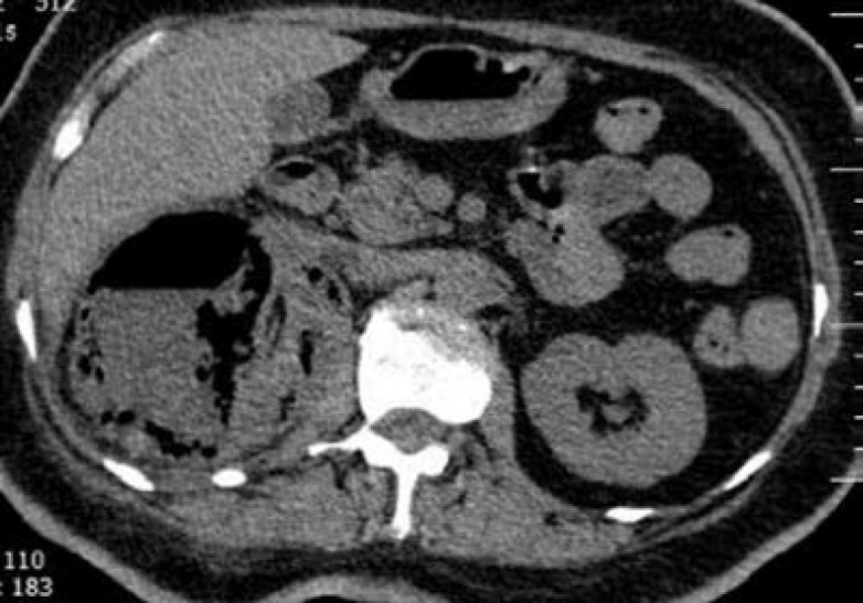
Case presentation: A 55–year-old diabetic woman presented to the emergency department with a complaint of nausea, vomiting, right upper abdominal pain, and fever with chills since 10 days. She revealed a 10-year history of poorly controlled diabetes on oral agent and kidney stones. On examination, the patient was found to be febrile (39 ℃) with tenderness in the right renal angle. Laboratory data has revealed the following findings: blood sugar (BS: 480 mg/dl), HbA1C: 13%, complete blood count (white blood cells (WBC): 13,900; polymorphonuclear leukocytes (PMN): 80%; lymphocytes: 18%; hemoglobin: 12 g/dl; and platelet: 118,000), blood urea nitrogen (BUN): 79 mg/dl, creatinine (Cr): 2.3 mg/dl, and erythrocyte sedimentation rate (ESR): 103 mm in 1 h. The urine analysis revealed 12–13 WBCs, 7–8 red blood cells (RBCs), and several bacteria. Urgent ultrasound indicated a heterogeneous mass in with focal echoes suggesting intraparenchymal gas, along with gross hydronephrosis and numerous stones, in the right kidney. The patient was treated with hydration, insulin, and intravenous imipenem 500 mg twice daily (adjusted with her creatinine). After 48 h, blood culture report was negative, whereas urine culture revealed presence of imipenem sensitive Citrobacter. Computed tomography (CT) scan without contrast indicated an enlarged, edematous right kidney with multiple air bubbles and air fluid levels. Based on the clinical and radiological findings, diagnosis was confirmed and right urgent nephrectomy was performed after 36 h of admission. The histopathology of the removed kidney revealed acute or chronic inflammation and necrosis, extending to the perinephric fat. The patient was discharged without any major complication after a 14-day hospital stay.
Learning points: Emphysematous pyelonephritis (EPN) is an acute, severe, and gas producing necrotizing bacterial infection that affects the renal parenchymal and surrounding tissues. The predisposing factors include: diabetes mellitus, urinary tract obstructions, and immune incompetence. Diabetes mellitus is the most commonly associated factor and up to 90% of the patients report uncontrolled diabetes mellitus. Bilateral renal involvement and obstruction has been observed in 5% and 30% of the patients, respectively. The most common pathogen causing EPN is Escherichia coli. Other pathogens have been reported including Klebsiella pneumoniae, Proteus mirabilis, and Pseudomonas aeruginosa. Several factors contribute in the pathogenesis of EPN including high levels of glucose inside the tissues, gas forming bacterial infection, impaired vascular blood supply, reduced host immunity, and obstruction in the urinary system. Clinical manifestations are similar to acute pyelonephritis, including fever, nausea, vomiting, and flank pain; however, often they do not respond to the medical treatment. Laboratory investigations often reveal leukocytosis with a shift to the left, thrombocytopenia, and elevation of the serum creatinine levels. As aforementioned, urine analysis reveals WBCs, RBCs, and several bacteria. The diagnosis is confirmed by radiological imaging. A plain abdominal X-ray can be more specific than the ultrasound, indicating the presence of gas in the kidney. The gold standard is abdominal CT scan that reveals the presence of gas and obstruction in the urinary tract systems. Treatment should commence with fluid resuscitation, antibiotic therapy, and control of blood sugar and electrolytes. Percutaneous drainage or DJ-stenting is recommended in the patients with urinary tract obstruction. If the aforementioned measures fail, then emergency nephrectomy should be considered.


 分享
分享

 求助内容:
求助内容: 应助结果提醒方式:
 扫码关注我们
