Jeffrey B Walker, Justin Loloi, Alexander Birk, Jay D Raman
{"title":"Computed Tomography Imaging Characteristics of Histologically Confirmed Papillary Renal Cell Carcinoma-Implications for Ancillary Imaging.","authors":"Jeffrey B Walker, Justin Loloi, Alexander Birk, Jay D Raman","doi":"10.15586/jkcvhl.2019.124","DOIUrl":null,"url":null,"abstract":"<p><p>Low-attenuation renal lesions on non-contrast computed tomography (CT) are often considered to be benign cysts without need for further imaging. However, the papillary subtype of renal cell carcinoma (RCC) may have similar radiographic characteristics. A single-center retrospective review was therefore performed to identify extirpated papillary RCC (pRCC) specimens with correlation made to preoperative tumor imaging characteristics. A total of 108 pRCC specimens were identified of which 84 (27 type I, 17 type 2, 40 unspecified) had CT imaging available for review. Non-contrast CT was available for 73 tumors with 16 (22%) demonstrating Hounsfield units (HU) measurements fewer than 20 at baseline without differences between papillary subtypes. Mean attenuation following contrast administration was similar between papillary subtypes (45 HU for type 1 pRCC and 49 HU for type 2). This study highlights that pathologically proven pRCC is a heterogeneous entity in terms of density on preoperative CT imaging. A non-contrast CT scan with HU fewer than 20 may not be an adequate evaluation for incidental renal masses, as over 1 in 5 pRCCs demonstrate lower attenuation than this cutoff. Further study is needed to identify the appropriate role of ancillary imaging in the workup of seemingly benign-appearing renal lesions.</p>","PeriodicalId":44291,"journal":{"name":"Journal of Kidney Cancer and VHL","volume":"6 2","pages":"10-14"},"PeriodicalIF":1.9000,"publicationDate":"2019-12-30","publicationTypes":"Journal Article","fieldsOfStudy":null,"isOpenAccess":false,"openAccessPdf":"https://www.ncbi.nlm.nih.gov/pmc/articles/PMC6942253/pdf/","citationCount":"5","resultStr":null,"platform":"Semanticscholar","paperid":null,"PeriodicalName":"Journal of Kidney Cancer and VHL","FirstCategoryId":"1085","ListUrlMain":"https://doi.org/10.15586/jkcvhl.2019.124","RegionNum":0,"RegionCategory":null,"ArticlePicture":[],"TitleCN":null,"AbstractTextCN":null,"PMCID":null,"EPubDate":"2019/1/1 0:00:00","PubModel":"eCollection","JCR":"Q3","JCRName":"ONCOLOGY","Score":null,"Total":0}
引用次数: 5
Abstract
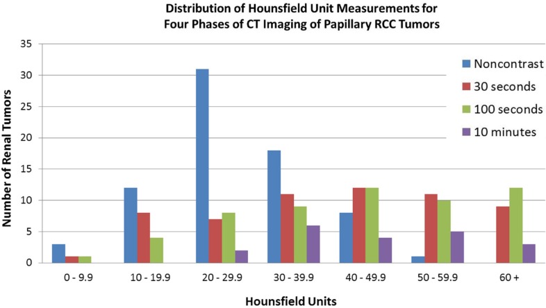
Low-attenuation renal lesions on non-contrast computed tomography (CT) are often considered to be benign cysts without need for further imaging. However, the papillary subtype of renal cell carcinoma (RCC) may have similar radiographic characteristics. A single-center retrospective review was therefore performed to identify extirpated papillary RCC (pRCC) specimens with correlation made to preoperative tumor imaging characteristics. A total of 108 pRCC specimens were identified of which 84 (27 type I, 17 type 2, 40 unspecified) had CT imaging available for review. Non-contrast CT was available for 73 tumors with 16 (22%) demonstrating Hounsfield units (HU) measurements fewer than 20 at baseline without differences between papillary subtypes. Mean attenuation following contrast administration was similar between papillary subtypes (45 HU for type 1 pRCC and 49 HU for type 2). This study highlights that pathologically proven pRCC is a heterogeneous entity in terms of density on preoperative CT imaging. A non-contrast CT scan with HU fewer than 20 may not be an adequate evaluation for incidental renal masses, as over 1 in 5 pRCCs demonstrate lower attenuation than this cutoff. Further study is needed to identify the appropriate role of ancillary imaging in the workup of seemingly benign-appearing renal lesions.


 分享
分享
 求助内容:
求助内容: 应助结果提醒方式:
 扫码关注我们
