Assessment of the Depth of Clinically Detected Approximal Caries Lesions Using Digital Imaging Fiber-Optic Transillumination in Comparison to Periapical Radiographs.
{"title":"Assessment of the Depth of Clinically Detected Approximal Caries Lesions Using Digital Imaging Fiber-Optic Transillumination in Comparison to Periapical Radiographs.","authors":"Auste Antipoviene, Monika Girijotaite, Egle Aida Bendoraitiene","doi":"10.5037/jomr.2020.11103","DOIUrl":null,"url":null,"abstract":"<p><strong>Objectives: </strong>The aim of present prospective clinical trial was to assess the depth of clinically detected approximal caries lesions using digital imaging fiber-optic transillumination in comparison to periapical radiographs.</p><p><strong>Material and methods: </strong>One calibrated examiner diagnosed 31 approximal carious lesions in 10 patients with a mean age of 21.8 (SD 1.1) years. The lesions were assessed using digital imaging fiber-optic transillumination (DIFOTI) and digital periapical radiographs (PA). Depending on the depth of the lesions, scores for demineralisation in PA (R) and DIFOTI (F) images were given by two examiners: R0/F0 - no demineralisation, R1/F1 - demineralisation confined to the outer half of the enamel, R2/F2 - into the inner half of the enamel, 3/3 - along the amelodentinal junction, R3/F3 - into the outer half of dentine, R4/F4 - into the inner part of the dentine. Spearman's rank correlation coefficient and kappa were calculated.</p><p><strong>Results: </strong>Spearman's rank correlation coefficient between DIFOTI and PA was 0.031 (P > 0.05), kappa was 0.077. In 26% of the cases, DIFOTI showed higher scores of demineralisation, relative to PA. In 36% of the cases, PA showed higher scores of demineralisation than DIFOTI. PA showed demineralisation into the outer and inner half of the dentine in 89% of the cases with underlying shadow and in 70% of the cases with opacities.</p><p><strong>Conclusions: </strong>Digital imaging fibre optic transillumination and periapical radiographs produce evaluations of the depth of approximal caries lesions that do not match.</p>","PeriodicalId":230885,"journal":{"name":"Journal of Oral & Maxillofacial Research","volume":"11 1","pages":"e3"},"PeriodicalIF":0.0000,"publicationDate":"2020-03-31","publicationTypes":"Journal Article","fieldsOfStudy":null,"isOpenAccess":false,"openAccessPdf":"https://ftp.ncbi.nlm.nih.gov/pub/pmc/oa_pdf/33/1e/jomr-11-e3.PMC7191380.pdf","citationCount":"1","resultStr":null,"platform":"Semanticscholar","paperid":null,"PeriodicalName":"Journal of Oral & Maxillofacial Research","FirstCategoryId":"1085","ListUrlMain":"https://doi.org/10.5037/jomr.2020.11103","RegionNum":0,"RegionCategory":null,"ArticlePicture":[],"TitleCN":null,"AbstractTextCN":null,"PMCID":null,"EPubDate":"2020/1/1 0:00:00","PubModel":"eCollection","JCR":"","JCRName":"","Score":null,"Total":0}
引用次数: 1
Abstract
Objectives: The aim of present prospective clinical trial was to assess the depth of clinically detected approximal caries lesions using digital imaging fiber-optic transillumination in comparison to periapical radiographs.
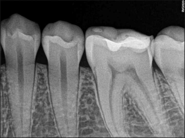
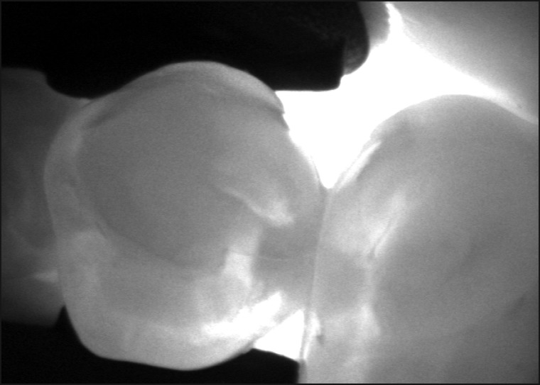
Material and methods: One calibrated examiner diagnosed 31 approximal carious lesions in 10 patients with a mean age of 21.8 (SD 1.1) years. The lesions were assessed using digital imaging fiber-optic transillumination (DIFOTI) and digital periapical radiographs (PA). Depending on the depth of the lesions, scores for demineralisation in PA (R) and DIFOTI (F) images were given by two examiners: R0/F0 - no demineralisation, R1/F1 - demineralisation confined to the outer half of the enamel, R2/F2 - into the inner half of the enamel, 3/3 - along the amelodentinal junction, R3/F3 - into the outer half of dentine, R4/F4 - into the inner part of the dentine. Spearman's rank correlation coefficient and kappa were calculated.
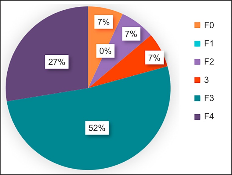
Results: Spearman's rank correlation coefficient between DIFOTI and PA was 0.031 (P > 0.05), kappa was 0.077. In 26% of the cases, DIFOTI showed higher scores of demineralisation, relative to PA. In 36% of the cases, PA showed higher scores of demineralisation than DIFOTI. PA showed demineralisation into the outer and inner half of the dentine in 89% of the cases with underlying shadow and in 70% of the cases with opacities.
Conclusions: Digital imaging fibre optic transillumination and periapical radiographs produce evaluations of the depth of approximal caries lesions that do not match.


 分享
分享

 求助内容:
求助内容: 应助结果提醒方式:
 扫码关注我们
