Proteomic investigation of protein adsorption to cerebral microdialysis membranes in surgically treated intracerebral hemorrhage patients - a pilot study.
Lovisa Tobieson, Zita Czifra, Karin Wåhlén, Niklas Marklund, Bijar Ghafouri
{"title":"Proteomic investigation of protein adsorption to cerebral microdialysis membranes in surgically treated intracerebral hemorrhage patients - a pilot study.","authors":"Lovisa Tobieson, Zita Czifra, Karin Wåhlén, Niklas Marklund, Bijar Ghafouri","doi":"10.1186/s12953-020-00163-7","DOIUrl":null,"url":null,"abstract":"<p><strong>Background: </strong>Cerebral microdialysis (CMD) is a minimally invasive technique for sampling the interstitial fluid in human brain tissue. CMD allows monitoring the metabolic state of tissue, as well as sampling macromolecules such as proteins and peptides. Recovery of proteins or peptides can be hampered by their adsorption to the CMD membrane as has been previously shown in-vitro<i>,</i> however, protein adsorption to CMD membranes has not been characterized following implantation in human brain tissue.</p><p><strong>Methods: </strong>In this paper, we describe the pattern of proteins adsorbed to CMD membranes compared to that of the microdialysate and of cerebrospinal fluid (CSF). We retrieved CMD membranes from three surgically treated intracerebral hemorrhage (ICH) patients, and analyzed protein adsorption to the membranes using two-dimensional gel electrophoresis (2-DE) in combination with nano-liquid mass spectrometry. We compared the proteome profile of three compartments; the CMD membrane, the microdialysate and ventricular CSF collected at time of CMD removal.</p><p><strong>Results: </strong>We found unique protein patterns in the molecular weight range of 10-35 kDa for each of the three compartments.</p><p><strong>Conclusion: </strong>This study highlights the importance of analyzing the membranes in addition to the microdialysate when using CMD to sample proteins for biomarker investigation.</p>","PeriodicalId":20857,"journal":{"name":"Proteome Science","volume":"18 ","pages":"7"},"PeriodicalIF":1.6000,"publicationDate":"2020-07-25","publicationTypes":"Journal Article","fieldsOfStudy":null,"isOpenAccess":false,"openAccessPdf":"https://sci-hub-pdf.com/10.1186/s12953-020-00163-7","citationCount":"2","resultStr":null,"platform":"Semanticscholar","paperid":null,"PeriodicalName":"Proteome Science","FirstCategoryId":"99","ListUrlMain":"https://doi.org/10.1186/s12953-020-00163-7","RegionNum":3,"RegionCategory":"生物学","ArticlePicture":[],"TitleCN":null,"AbstractTextCN":null,"PMCID":null,"EPubDate":"2020/1/1 0:00:00","PubModel":"eCollection","JCR":"Q3","JCRName":"BIOCHEMICAL RESEARCH METHODS","Score":null,"Total":0}
引用次数: 2
Abstract
Background: Cerebral microdialysis (CMD) is a minimally invasive technique for sampling the interstitial fluid in human brain tissue. CMD allows monitoring the metabolic state of tissue, as well as sampling macromolecules such as proteins and peptides. Recovery of proteins or peptides can be hampered by their adsorption to the CMD membrane as has been previously shown in-vitro, however, protein adsorption to CMD membranes has not been characterized following implantation in human brain tissue.
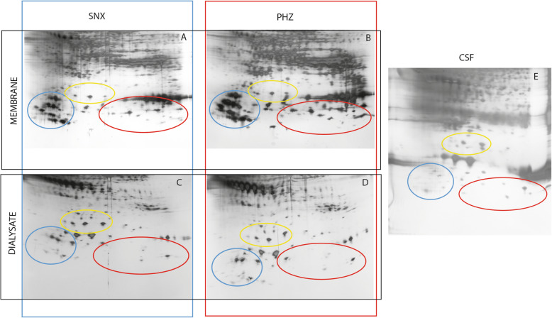
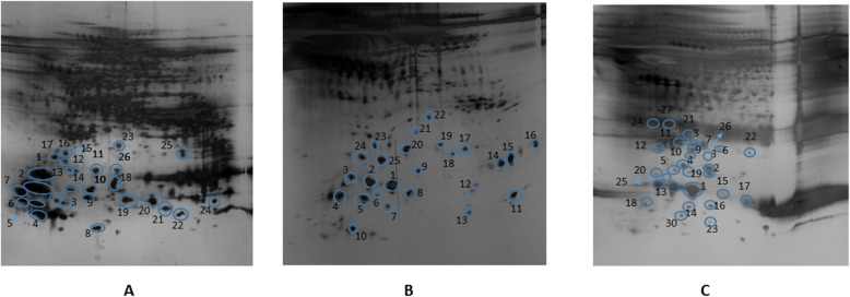
Methods: In this paper, we describe the pattern of proteins adsorbed to CMD membranes compared to that of the microdialysate and of cerebrospinal fluid (CSF). We retrieved CMD membranes from three surgically treated intracerebral hemorrhage (ICH) patients, and analyzed protein adsorption to the membranes using two-dimensional gel electrophoresis (2-DE) in combination with nano-liquid mass spectrometry. We compared the proteome profile of three compartments; the CMD membrane, the microdialysate and ventricular CSF collected at time of CMD removal.
Results: We found unique protein patterns in the molecular weight range of 10-35 kDa for each of the three compartments.
Conclusion: This study highlights the importance of analyzing the membranes in addition to the microdialysate when using CMD to sample proteins for biomarker investigation.
期刊介绍:
Proteome Science is an open access journal publishing research in the area of systems studies. Proteome Science considers manuscripts based on all aspects of functional and structural proteomics, genomics, metabolomics, systems analysis and metabiome analysis. It encourages the submissions of studies that use large-scale or systems analysis of biomolecules in a cellular, organismal and/or environmental context.
Studies that describe novel biological or clinical insights as well as methods-focused studies that describe novel methods for the large-scale study of any and all biomolecules in cells and tissues, such as mass spectrometry, protein and nucleic acid microarrays, genomics, next-generation sequencing and computational algorithms and methods are all within the scope of Proteome Science, as are electron topography, structural methods, proteogenomics, chemical proteomics, stem cell proteomics, organelle proteomics, plant and microbial proteomics.
In spite of its name, Proteome Science considers all aspects of large-scale and systems studies because ultimately any mechanism that results in genomic and metabolomic changes will affect or be affected by the proteome. To reflect this intrinsic relationship of biological systems, Proteome Science will consider all such articles.


 分享
分享
 求助内容:
求助内容: 应助结果提醒方式:
 扫码关注我们
