Liang Han, Jose Ronaldo Lima de Carvalho, Rait Parmann, Tongalp H Tezel, Stanley Chang, Tarun Sharma, Janet R Sparrow
{"title":"Central Serous Chorioretinopathy Analyzed by Multimodal Imaging.","authors":"Liang Han, Jose Ronaldo Lima de Carvalho, Rait Parmann, Tongalp H Tezel, Stanley Chang, Tarun Sharma, Janet R Sparrow","doi":"10.1167/tvst.10.1.15","DOIUrl":null,"url":null,"abstract":"<p><strong>Purpose: </strong>We correlated quantitative fundus autofluorescence (qAF) with other fundus features in patients exhibiting central serous chorioretinopathy (CSC).</p><p><strong>Methods: </strong>Short wavelength fundus autofluorescence (SW-AF, 488 nm excitation) was measured by qAF. Using nonnormalized images qAF values were calculated within eight concentric segments (qAF<sub>8</sub>) located at an eccentricity of 7° to 9°. Horizontal spectral domain optical coherence tomography (SD-OCT) scans and near-infrared fundus autofluorescence images (NIR-AF) were studied.</p><p><strong>Results: </strong>Thirty-six eyes of 20 patients (mean age 48.7± 8.5 years) diagnosed with CSC were studied. Thirteen patients had bilateral disease; four patients were female. In 22 eyes CSC was present in the macula; in one eye the lesion was in a peripapillary location, 10 involved both locations, and three were unaffected. Serous retinal detachment, retinal pigmented epithelial detachment (PED), outer retinal atrophy and subRPE hypertransmission were all features identifiable by SD-OCT. NIR-AF images were helpful in detecting foveal and parafoveal lesions. Sampling for retina-wide elevations in SW-AF intensity by measuring qAF<sub>8</sub> did not indicate a generalizable relationship amongst CSC-diagnosed eyes. However, color-coded qAF images revealed alterations in SW-AF topography and intensity relative to healthy eyes at the same locations. Thus zones of higher than normal qAF intensity were found in association with SD-OCT detectable PED; loss of ellipsoid zone and interdigitation zone; and hyperreflectivity in outer retina. Pronounced decreases in qAF colocalized with serous retinal detachment and with outer retinal degeneration that included hypertransmission of SD-OCT signal into the choroid.</p><p><strong>Conclusions: </strong>Localized elevations in qAF reflect increased bisretinoid in association with CSC lesions.</p><p><strong>Translational relevance: </strong>Foci of elevated qAF at some stages of CSC contribute to the natural history of the disease.</p>","PeriodicalId":23322,"journal":{"name":"Translational Vision Science & Technology","volume":" ","pages":"15"},"PeriodicalIF":2.6000,"publicationDate":"2021-01-08","publicationTypes":"Journal Article","fieldsOfStudy":null,"isOpenAccess":false,"openAccessPdf":"https://ftp.ncbi.nlm.nih.gov/pub/pmc/oa_pdf/e3/f9/tvst-10-1-15.PMC7804571.pdf","citationCount":"6","resultStr":null,"platform":"Semanticscholar","paperid":null,"PeriodicalName":"Translational Vision Science & Technology","FirstCategoryId":"3","ListUrlMain":"https://doi.org/10.1167/tvst.10.1.15","RegionNum":3,"RegionCategory":"医学","ArticlePicture":[],"TitleCN":null,"AbstractTextCN":null,"PMCID":null,"EPubDate":"2021/1/1 0:00:00","PubModel":"eCollection","JCR":"Q2","JCRName":"OPHTHALMOLOGY","Score":null,"Total":0}
引用次数: 6
Abstract
Purpose: We correlated quantitative fundus autofluorescence (qAF) with other fundus features in patients exhibiting central serous chorioretinopathy (CSC).
Methods: Short wavelength fundus autofluorescence (SW-AF, 488 nm excitation) was measured by qAF. Using nonnormalized images qAF values were calculated within eight concentric segments (qAF8) located at an eccentricity of 7° to 9°. Horizontal spectral domain optical coherence tomography (SD-OCT) scans and near-infrared fundus autofluorescence images (NIR-AF) were studied.
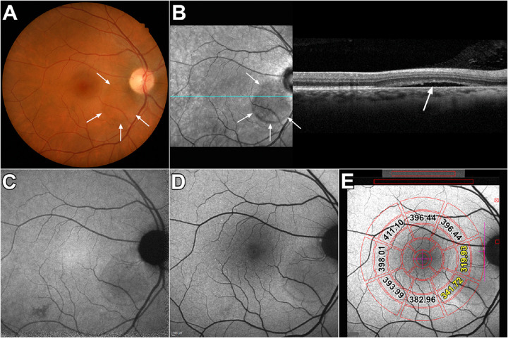
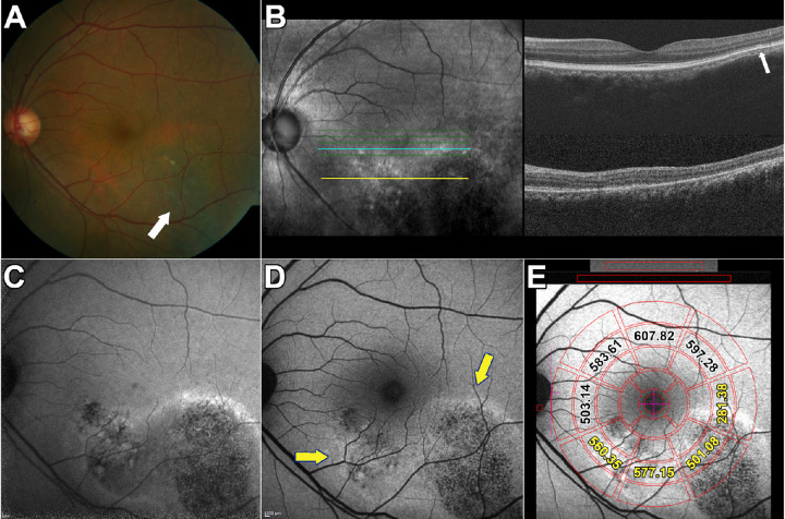
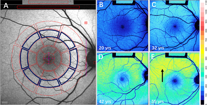
Results: Thirty-six eyes of 20 patients (mean age 48.7± 8.5 years) diagnosed with CSC were studied. Thirteen patients had bilateral disease; four patients were female. In 22 eyes CSC was present in the macula; in one eye the lesion was in a peripapillary location, 10 involved both locations, and three were unaffected. Serous retinal detachment, retinal pigmented epithelial detachment (PED), outer retinal atrophy and subRPE hypertransmission were all features identifiable by SD-OCT. NIR-AF images were helpful in detecting foveal and parafoveal lesions. Sampling for retina-wide elevations in SW-AF intensity by measuring qAF8 did not indicate a generalizable relationship amongst CSC-diagnosed eyes. However, color-coded qAF images revealed alterations in SW-AF topography and intensity relative to healthy eyes at the same locations. Thus zones of higher than normal qAF intensity were found in association with SD-OCT detectable PED; loss of ellipsoid zone and interdigitation zone; and hyperreflectivity in outer retina. Pronounced decreases in qAF colocalized with serous retinal detachment and with outer retinal degeneration that included hypertransmission of SD-OCT signal into the choroid.
Conclusions: Localized elevations in qAF reflect increased bisretinoid in association with CSC lesions.
Translational relevance: Foci of elevated qAF at some stages of CSC contribute to the natural history of the disease.
期刊介绍:
Translational Vision Science & Technology (TVST), an official journal of the Association for Research in Vision and Ophthalmology (ARVO), an international organization whose purpose is to advance research worldwide into understanding the visual system and preventing, treating and curing its disorders, is an online, open access, peer-reviewed journal emphasizing multidisciplinary research that bridges the gap between basic research and clinical care. A highly qualified and diverse group of Associate Editors and Editorial Board Members is led by Editor-in-Chief Marco Zarbin, MD, PhD, FARVO.
The journal covers a broad spectrum of work, including but not limited to:
Applications of stem cell technology for regenerative medicine,
Development of new animal models of human diseases,
Tissue bioengineering,
Chemical engineering to improve virus-based gene delivery,
Nanotechnology for drug delivery,
Design and synthesis of artificial extracellular matrices,
Development of a true microsurgical operating environment,
Refining data analysis algorithms to improve in vivo imaging technology,
Results of Phase 1 clinical trials,
Reverse translational ("bedside to bench") research.
TVST seeks manuscripts from scientists and clinicians with diverse backgrounds ranging from basic chemistry to ophthalmic surgery that will advance or change the way we understand and/or treat vision-threatening diseases. TVST encourages the use of color, multimedia, hyperlinks, program code and other digital enhancements.


 分享
分享
 求助内容:
求助内容: 应助结果提醒方式:
 扫码关注我们
