{"title":"Raman Spectroscopic Assessment of Myocardial Viability in Langendorff-Perfused Ischemic Rat Hearts.","authors":"Koki Ikemoto, Kosuke Hashimoto, Yoshinori Harada, Yasuaki Kumamoto, Michiyo Hayakawa, Kentaro Mochizuki, Kazuhiko Matsuo, Kenta Yashiro, Hitoshi Yaku, Tetsuro Takamatsu, Hideo Tanaka","doi":"10.1267/ahc.21-00016","DOIUrl":null,"url":null,"abstract":"<p><p>Spontaneous Raman spectroscopy, which senses changes in cellular contents of reduced cytochrome c, could be a powerful tool for label-free evaluation of ischemic hearts. However, undetermined is whether it is applicable to evaluation of myocardial viability in ischemic hearts. To address this issue, we investigated sequential changes in Raman spectra of the subepicardial myocardium in the Langendorff-perfused rat heart before and during ligation of the left coronary artery and its subsequent release and re-ligation. Under 532-nm wavelength excitation, the Raman peak intensity of reduced cytochrome c at 747 cm<sup>-1</sup> increased quickly after the coronary ligation, and reached a quasi-steady state within 30 min. Subsequent reperfusion of the heart after a short-term (30-min) ligation that simulates reversible conditions resulted in quick recovery of the peak intensity to the baseline. Further re-ligation resulted in resurgence of the peak intensity to nearly the identical value to the first ischemia value. In contrast, reperfusion after prolonged (120-min) ligation that assumes irreversible states resulted in incomplete recovery of the peak intensity, and re-ligation resulted in inadequate resurgence. Electron microscopic observations confirmed the spectral findings. Together, the Raman spectroscopic measurement for cytochrome c could be applicable to evaluation of viability of the ischemic myocardium without labeling.</p>","PeriodicalId":6888,"journal":{"name":"Acta Histochemica Et Cytochemica","volume":"54 2","pages":"65-72"},"PeriodicalIF":1.8000,"publicationDate":"2021-04-28","publicationTypes":"Journal Article","fieldsOfStudy":null,"isOpenAccess":false,"openAccessPdf":"https://ftp.ncbi.nlm.nih.gov/pub/pmc/oa_pdf/14/f7/ahc-054-65.PMC8116620.pdf","citationCount":"4","resultStr":null,"platform":"Semanticscholar","paperid":null,"PeriodicalName":"Acta Histochemica Et Cytochemica","FirstCategoryId":"99","ListUrlMain":"https://doi.org/10.1267/ahc.21-00016","RegionNum":4,"RegionCategory":"生物学","ArticlePicture":[],"TitleCN":null,"AbstractTextCN":null,"PMCID":null,"EPubDate":"2021/4/17 0:00:00","PubModel":"Epub","JCR":"Q4","JCRName":"CELL BIOLOGY","Score":null,"Total":0}
引用次数: 4
Abstract
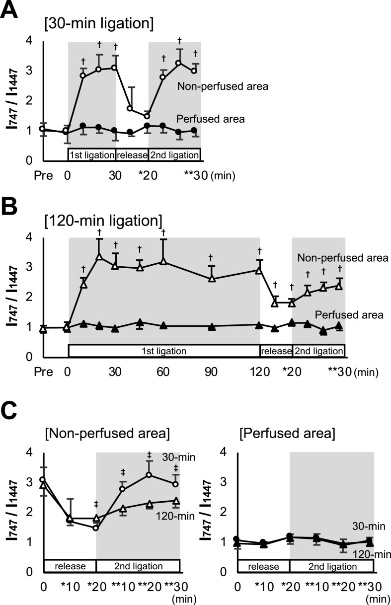
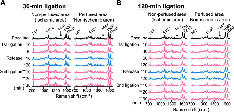
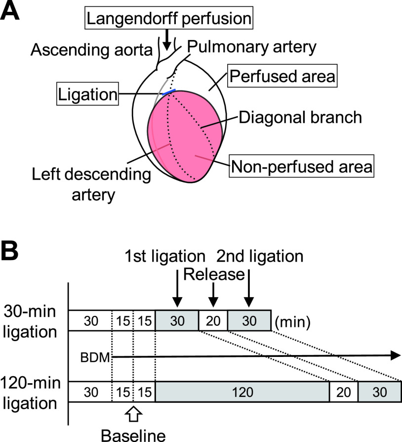
Spontaneous Raman spectroscopy, which senses changes in cellular contents of reduced cytochrome c, could be a powerful tool for label-free evaluation of ischemic hearts. However, undetermined is whether it is applicable to evaluation of myocardial viability in ischemic hearts. To address this issue, we investigated sequential changes in Raman spectra of the subepicardial myocardium in the Langendorff-perfused rat heart before and during ligation of the left coronary artery and its subsequent release and re-ligation. Under 532-nm wavelength excitation, the Raman peak intensity of reduced cytochrome c at 747 cm-1 increased quickly after the coronary ligation, and reached a quasi-steady state within 30 min. Subsequent reperfusion of the heart after a short-term (30-min) ligation that simulates reversible conditions resulted in quick recovery of the peak intensity to the baseline. Further re-ligation resulted in resurgence of the peak intensity to nearly the identical value to the first ischemia value. In contrast, reperfusion after prolonged (120-min) ligation that assumes irreversible states resulted in incomplete recovery of the peak intensity, and re-ligation resulted in inadequate resurgence. Electron microscopic observations confirmed the spectral findings. Together, the Raman spectroscopic measurement for cytochrome c could be applicable to evaluation of viability of the ischemic myocardium without labeling.
期刊介绍:
Acta Histochemica et Cytochemica is the official online journal of the Japan Society of Histochemistry and Cytochemistry. It is intended primarily for rapid publication of concise, original articles in the fields of histochemistry and cytochemistry. Manuscripts oriented towards methodological subjects that contain significant technical advances in these fields are also welcome. Manuscripts in English are accepted from investigators in any country, whether or not they are members of the Japan Society of Histochemistry and Cytochemistry. Manuscripts should be original work that has not been previously published and is not being considered for publication elsewhere, with the exception of abstracts. Manuscripts with essentially the same content as a paper that has been published or accepted, or is under consideration for publication, will not be considered. All submitted papers will be peer-reviewed by at least two referees selected by an appropriate Associate Editor. Acceptance is based on scientific significance, originality, and clarity. When required, a revised manuscript should be submitted within 3 months, otherwise it will be considered to be a new submission. The Editor-in-Chief will make all final decisions regarding acceptance.


 分享
分享
 求助内容:
求助内容: 应助结果提醒方式:
 扫码关注我们
