Elisabeth Kugler, Ryan Snodgrass, George Bowley, Karen Plant, Jovana Serbanovic-Canic, Noémie Hamilton, Paul C Evans, Timothy Chico, Paul Armitage
{"title":"The effect of absent blood flow on the zebrafish cerebral and trunk vasculature.","authors":"Elisabeth Kugler, Ryan Snodgrass, George Bowley, Karen Plant, Jovana Serbanovic-Canic, Noémie Hamilton, Paul C Evans, Timothy Chico, Paul Armitage","doi":"10.1530/VB-21-0009","DOIUrl":null,"url":null,"abstract":"<p><p>The role of blood flow in vascular development is complex and context-dependent. In this study, we quantify the effect of the lack of blood flow on embryonic vascular development on two vascular beds, namely the cerebral and trunk vasculature in zebrafish. We perform this by analysing vascular topology, endothelial cell (EC) number, EC distribution, apoptosis, and inflammatory response in animals with normal blood flow or absent blood flow. We find that absent blood flow reduced vascular area and EC number significantly in both examined vascular beds, but the effect is more severe in the cerebral vasculature, and severity increases over time. Absent blood flow leads to an increase in non-EC-specific apoptosis without increasing tissue inflammation, as quantified by cerebral immune cell numbers and nitric oxide. Similarly, while stereotypic vascular patterning in the trunk is maintained, intra-cerebral vessels show altered patterning, which is likely to be due to vessels failing to initiate effective fusion and anastomosis rather than sprouting or path-seeking. In conclusion, blood flow is essential for cellular survival in both the trunk and cerebral vasculature, but particularly intra-cerebral vessels are affected by the lack of blood flow, suggesting that responses to blood flow differ between these two vascular beds.</p>","PeriodicalId":75294,"journal":{"name":"Vascular biology (Bristol, England)","volume":"3 1","pages":"1-16"},"PeriodicalIF":0.0000,"publicationDate":"2021-07-29","publicationTypes":"Journal Article","fieldsOfStudy":null,"isOpenAccess":false,"openAccessPdf":"https://ftp.ncbi.nlm.nih.gov/pub/pmc/oa_pdf/48/93/VB-21-0009.PMC8428019.pdf","citationCount":"0","resultStr":null,"platform":"Semanticscholar","paperid":null,"PeriodicalName":"Vascular biology (Bristol, England)","FirstCategoryId":"1085","ListUrlMain":"https://doi.org/10.1530/VB-21-0009","RegionNum":0,"RegionCategory":null,"ArticlePicture":[],"TitleCN":null,"AbstractTextCN":null,"PMCID":null,"EPubDate":"2021/1/1 0:00:00","PubModel":"eCollection","JCR":"","JCRName":"","Score":null,"Total":0}
引用次数: 0
Abstract
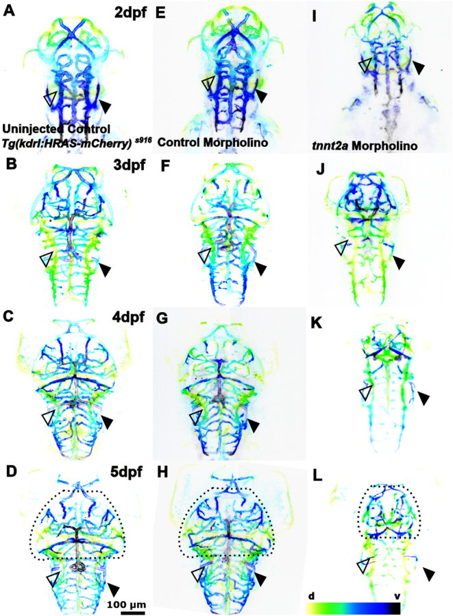
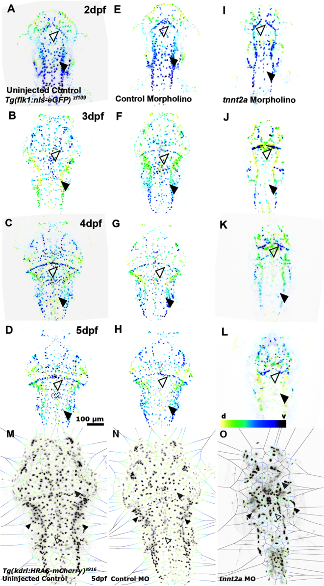
The role of blood flow in vascular development is complex and context-dependent. In this study, we quantify the effect of the lack of blood flow on embryonic vascular development on two vascular beds, namely the cerebral and trunk vasculature in zebrafish. We perform this by analysing vascular topology, endothelial cell (EC) number, EC distribution, apoptosis, and inflammatory response in animals with normal blood flow or absent blood flow. We find that absent blood flow reduced vascular area and EC number significantly in both examined vascular beds, but the effect is more severe in the cerebral vasculature, and severity increases over time. Absent blood flow leads to an increase in non-EC-specific apoptosis without increasing tissue inflammation, as quantified by cerebral immune cell numbers and nitric oxide. Similarly, while stereotypic vascular patterning in the trunk is maintained, intra-cerebral vessels show altered patterning, which is likely to be due to vessels failing to initiate effective fusion and anastomosis rather than sprouting or path-seeking. In conclusion, blood flow is essential for cellular survival in both the trunk and cerebral vasculature, but particularly intra-cerebral vessels are affected by the lack of blood flow, suggesting that responses to blood flow differ between these two vascular beds.


 分享
分享

 求助内容:
求助内容: 应助结果提醒方式:
 扫码关注我们
