{"title":"Muscle volume evaluation using 3DCT for congenital clubfoot.","authors":"Masataka Kakihana, Yuki Tochigi, Satoru Ozeki, Tetsuya Jinno","doi":"10.1177/20584601211062084","DOIUrl":null,"url":null,"abstract":"<p><strong>Background: </strong>In congenital clubfoot, the lower leg is very thin and the calf muscles are hypoplasic. However, there are few studies reporting real muscle volume.</p><p><strong>Purpose: </strong>The purpose of this study is to assay the muscle volume in congenital clubfoot using 3DCT and to quantify the degree of the hypoplasia.</p><p><strong>Material and methods: </strong>From January 2015 to December 2016, nine consecutive patients, seven male and two female, with unilateral congenital clubfeet were recruited for CT scans. Axial transverse sectional CT scans were acquired from the delineation of the fibular head to the tibial plafond. From the data, we rendered the entire muscle in 3D for muscle volume assay, and further segmented the posterior musculature for comparison between the normal and affected sides.</p><p><strong>Results: </strong>The whole muscle volume on the normal side was 291.23 cm<sup>3</sup> (181.23-593.49) and that on the affected side was 225.08 cm<sup>3</sup> (120.71-429.08), for an affected side to normal side ratio of 0.79 (0.72-0.9), which was significantly smaller (<i>p <</i> .01). Posterior muscle volume on the normal side was 175.81 cm<sup>3</sup> (103.72-376.32) and that on the affected side was 106.52 cm<sup>3</sup> (58.3-188.39). The ratio of posterior muscle to whole muscle on the normal side was 0.62 (0.46-0.75), and that on the affected side was 0.48 (0.4-0.55), such that the affected side was significantly smaller (<i>p <</i> .01).</p><p><strong>Conclusion: </strong>This study contributes quantitative data supporting the longstanding observations that the posterior calf muscles are significantly smaller on the affected side compared to the normal side in congenital clubfoot, and further underscores the importance of the extending the excursion of these muscles.</p>","PeriodicalId":72063,"journal":{"name":"Acta radiologica open","volume":"10 11","pages":"20584601211062084"},"PeriodicalIF":1.0000,"publicationDate":"2021-12-03","publicationTypes":"Journal Article","fieldsOfStudy":null,"isOpenAccess":false,"openAccessPdf":"https://ftp.ncbi.nlm.nih.gov/pub/pmc/oa_pdf/73/01/10.1177_20584601211062084.PMC8646796.pdf","citationCount":"1","resultStr":null,"platform":"Semanticscholar","paperid":null,"PeriodicalName":"Acta radiologica open","FirstCategoryId":"1085","ListUrlMain":"https://doi.org/10.1177/20584601211062084","RegionNum":0,"RegionCategory":null,"ArticlePicture":[],"TitleCN":null,"AbstractTextCN":null,"PMCID":null,"EPubDate":"2021/11/1 0:00:00","PubModel":"eCollection","JCR":"Q4","JCRName":"RADIOLOGY, NUCLEAR MEDICINE & MEDICAL IMAGING","Score":null,"Total":0}
引用次数: 1
Abstract
Background: In congenital clubfoot, the lower leg is very thin and the calf muscles are hypoplasic. However, there are few studies reporting real muscle volume.
Purpose: The purpose of this study is to assay the muscle volume in congenital clubfoot using 3DCT and to quantify the degree of the hypoplasia.
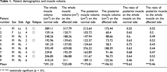
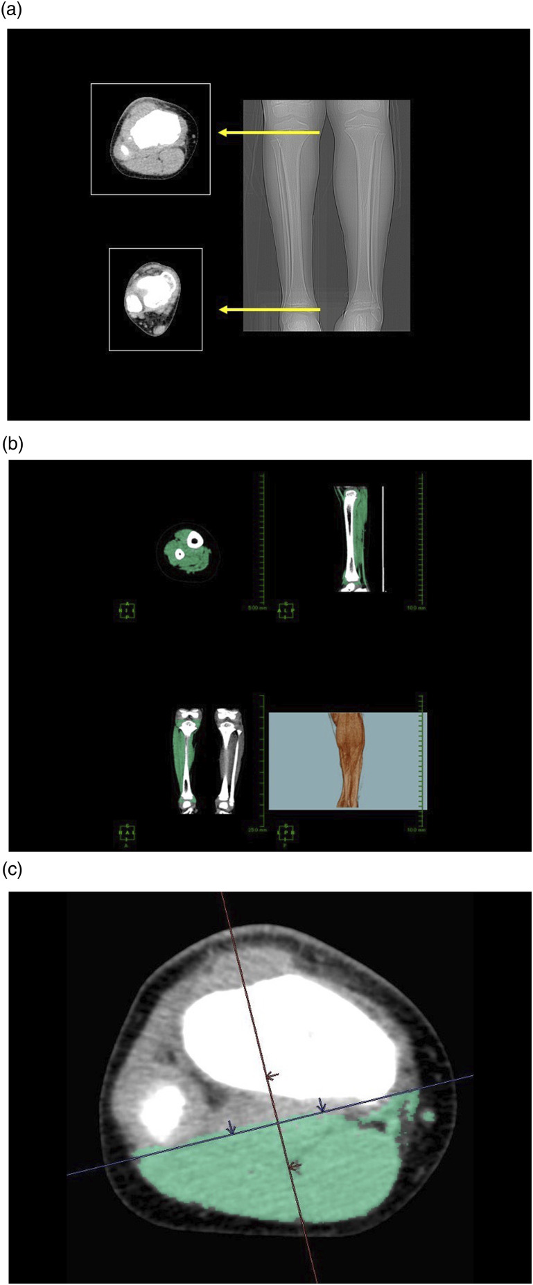
Material and methods: From January 2015 to December 2016, nine consecutive patients, seven male and two female, with unilateral congenital clubfeet were recruited for CT scans. Axial transverse sectional CT scans were acquired from the delineation of the fibular head to the tibial plafond. From the data, we rendered the entire muscle in 3D for muscle volume assay, and further segmented the posterior musculature for comparison between the normal and affected sides.
Results: The whole muscle volume on the normal side was 291.23 cm3 (181.23-593.49) and that on the affected side was 225.08 cm3 (120.71-429.08), for an affected side to normal side ratio of 0.79 (0.72-0.9), which was significantly smaller (p < .01). Posterior muscle volume on the normal side was 175.81 cm3 (103.72-376.32) and that on the affected side was 106.52 cm3 (58.3-188.39). The ratio of posterior muscle to whole muscle on the normal side was 0.62 (0.46-0.75), and that on the affected side was 0.48 (0.4-0.55), such that the affected side was significantly smaller (p < .01).
Conclusion: This study contributes quantitative data supporting the longstanding observations that the posterior calf muscles are significantly smaller on the affected side compared to the normal side in congenital clubfoot, and further underscores the importance of the extending the excursion of these muscles.


 分享
分享

 求助内容:
求助内容: 应助结果提醒方式:
 扫码关注我们
