{"title":"Ultrastructure of the fertilized egg envelope from Melanotaenia praecox, Melanotaeniidae, Teleostei","authors":"Joon Hyung Sohn, Dong Heui Kim","doi":"10.1186/s42649-021-00052-z","DOIUrl":null,"url":null,"abstract":"<p>We examined the morphology of fertilized egg and ultrastructures of fertilized egg envelopes of dwarf rainbowfish (<i>Melanotaenia praecox</i>) belong to Melanotaeniidae using light and electron microscopes. The fertilized eggs were spherical with adhesive filament, transparent, demersal, and had a narrow perivitelline space and small oil droplets. The size of fertilized egg was 1.02?±?0.18?mm (<i>n</i>?=?30), and there were two kinds of adhesive filament on the fertilized eggs. The long and thick (diameter 12.22?±?0.52?μm, <i>n</i>?=?20) adhesive filaments were only at the area of animal pole, and short and thin (diameter 1.99?±?0.23?μm, <i>n</i>?=?20) adhesive filaments were around the long filaments. A micropyle was conical shaped with adhesive filament and located near the animal pole of egg. The outer surface of fertilized egg was rough side. Also, the total thickness of the fertilized egg envelope was about 7.46?±?0.41?μm (<i>n</i>?=?20), the fertilized egg envelope consisted of two layers, an inner lamellae layer and an outer layer with high electron-density. And the inner layer was 8 layers. Collectively, these morphological characteristics and adhesive property of fertilized egg with adhesive filaments, and ultrastructures of micropyle, outer surface, and section of fertilized egg envelope are showed species specificity.</p>","PeriodicalId":470,"journal":{"name":"Applied Microscopy","volume":"51 1","pages":""},"PeriodicalIF":0.0000,"publicationDate":"2021-04-01","publicationTypes":"Journal Article","fieldsOfStudy":null,"isOpenAccess":false,"openAccessPdf":"https://sci-hub-pdf.com/10.1186/s42649-021-00052-z","citationCount":"1","resultStr":null,"platform":"Semanticscholar","paperid":null,"PeriodicalName":"Applied Microscopy","FirstCategoryId":"1085","ListUrlMain":"https://link.springer.com/article/10.1186/s42649-021-00052-z","RegionNum":0,"RegionCategory":null,"ArticlePicture":[],"TitleCN":null,"AbstractTextCN":null,"PMCID":null,"EPubDate":"","PubModel":"","JCR":"Q3","JCRName":"Immunology and Microbiology","Score":null,"Total":0}
引用次数: 1
Abstract
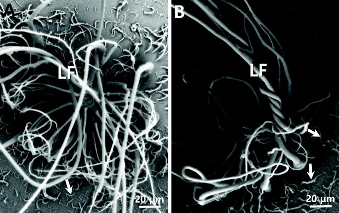
We examined the morphology of fertilized egg and ultrastructures of fertilized egg envelopes of dwarf rainbowfish (Melanotaenia praecox) belong to Melanotaeniidae using light and electron microscopes. The fertilized eggs were spherical with adhesive filament, transparent, demersal, and had a narrow perivitelline space and small oil droplets. The size of fertilized egg was 1.02?±?0.18?mm (n?=?30), and there were two kinds of adhesive filament on the fertilized eggs. The long and thick (diameter 12.22?±?0.52?μm, n?=?20) adhesive filaments were only at the area of animal pole, and short and thin (diameter 1.99?±?0.23?μm, n?=?20) adhesive filaments were around the long filaments. A micropyle was conical shaped with adhesive filament and located near the animal pole of egg. The outer surface of fertilized egg was rough side. Also, the total thickness of the fertilized egg envelope was about 7.46?±?0.41?μm (n?=?20), the fertilized egg envelope consisted of two layers, an inner lamellae layer and an outer layer with high electron-density. And the inner layer was 8 layers. Collectively, these morphological characteristics and adhesive property of fertilized egg with adhesive filaments, and ultrastructures of micropyle, outer surface, and section of fertilized egg envelope are showed species specificity.
Applied MicroscopyImmunology and Microbiology-Applied Microbiology and Biotechnology
CiteScore
3.40
自引率
0.00%
发文量
10
审稿时长
10 weeks
期刊介绍:
Applied Microscopy is a peer-reviewed journal sponsored by the Korean Society of Microscopy. The journal covers all the interdisciplinary fields of technological developments in new microscopy methods and instrumentation and their applications to biological or materials science for determining structure and chemistry. ISSN: 22875123, 22874445.


 分享
分享
 求助内容:
求助内容: 应助结果提醒方式:
 扫码关注我们
