Debdeep Banerjee, Laura L Magnelli, Mailin Oliva, Neha Malik, Brittney M Ginsburg, Joseph R Grajo, Tony S Brar, Donevan R Westerveld, Dennis Yang, Peter V Draganov
{"title":"Characterizing a CT esophagram protocol after flexible endoscopic diverticulotomy for Zenker's diverticulum: a retrospective series.","authors":"Debdeep Banerjee, Laura L Magnelli, Mailin Oliva, Neha Malik, Brittney M Ginsburg, Joseph R Grajo, Tony S Brar, Donevan R Westerveld, Dennis Yang, Peter V Draganov","doi":"10.21037/tgh-20-269","DOIUrl":null,"url":null,"abstract":"<p><strong>Background: </strong>Flexible endoscopic cricopharyngeal myotomy and septotomy offer a minimally invasive transluminal option for the treatment of symptomatic Zenker's diverticulum (ZD). There is currently no consensus regarding postoperative follow-up imaging. We suggest a standardized computed tomography (CT) esophagram protocol for radiographic evaluation of postoperative findings.</p><p><strong>Methods: </strong>Single center retrospective analysis of patients with symptomatic ZD who underwent flexible endoscopic diverticulotomy and postoperative imaging with CT esophagram from January 2015 to March 2020. An experienced radiologist blinded to the initial imaging reports prospectively interpreted all CT esophagram findings, in order to minimize bias.</p><p><strong>Results: </strong>Twenty-one patients underwent CT esophagram following flexible endoscopic diverticulotomy for ZD. Diverticulotomy was technically successful in all patients. Most common findings on imaging included: atelectasis (13/21; 62%), persistent esophageal diverticulum (7/21; 33%), pneumomediastinum (3/21; 14%), aspiration (2/21; 10%), and extraluminal air and contrast extravasation consistent with focal esophageal perforation (1/21; 5%).</p><p><strong>Conclusions: </strong>We describe a standardized, simple, and accessible CT esophagram protocol for postoperative imaging of patients with post-flexible endoscopic cricopharyngeal myotomy and septotomy for ZD. CT esophagram facilitates a definitive exclusion of focal esophageal perforation as a postoperative complication of flexible endoscopic diverticulotomy by ruling out extraluminal air and contrast extravasation.</p>","PeriodicalId":23267,"journal":{"name":"Translational gastroenterology and hepatology","volume":" ","pages":"34"},"PeriodicalIF":3.0000,"publicationDate":"2022-10-25","publicationTypes":"Journal Article","fieldsOfStudy":null,"isOpenAccess":false,"openAccessPdf":"https://ftp.ncbi.nlm.nih.gov/pub/pmc/oa_pdf/2d/1b/tgh-07-20-269.PMC9469010.pdf","citationCount":"0","resultStr":null,"platform":"Semanticscholar","paperid":null,"PeriodicalName":"Translational gastroenterology and hepatology","FirstCategoryId":"3","ListUrlMain":"https://doi.org/10.21037/tgh-20-269","RegionNum":4,"RegionCategory":"医学","ArticlePicture":[],"TitleCN":null,"AbstractTextCN":null,"PMCID":null,"EPubDate":"2022/1/1 0:00:00","PubModel":"eCollection","JCR":"Q1","JCRName":"Medicine","Score":null,"Total":0}
引用次数: 0
Abstract
Background: Flexible endoscopic cricopharyngeal myotomy and septotomy offer a minimally invasive transluminal option for the treatment of symptomatic Zenker's diverticulum (ZD). There is currently no consensus regarding postoperative follow-up imaging. We suggest a standardized computed tomography (CT) esophagram protocol for radiographic evaluation of postoperative findings.
Methods: Single center retrospective analysis of patients with symptomatic ZD who underwent flexible endoscopic diverticulotomy and postoperative imaging with CT esophagram from January 2015 to March 2020. An experienced radiologist blinded to the initial imaging reports prospectively interpreted all CT esophagram findings, in order to minimize bias.
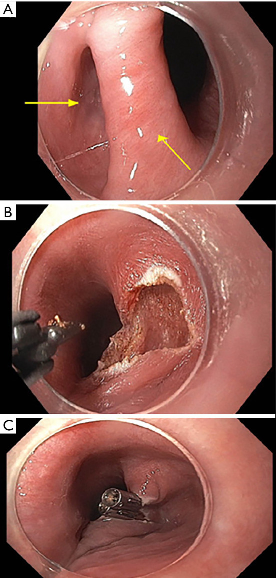
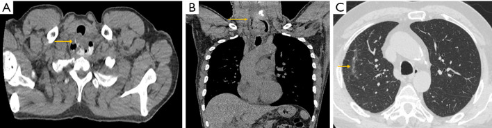
Results: Twenty-one patients underwent CT esophagram following flexible endoscopic diverticulotomy for ZD. Diverticulotomy was technically successful in all patients. Most common findings on imaging included: atelectasis (13/21; 62%), persistent esophageal diverticulum (7/21; 33%), pneumomediastinum (3/21; 14%), aspiration (2/21; 10%), and extraluminal air and contrast extravasation consistent with focal esophageal perforation (1/21; 5%).
Conclusions: We describe a standardized, simple, and accessible CT esophagram protocol for postoperative imaging of patients with post-flexible endoscopic cricopharyngeal myotomy and septotomy for ZD. CT esophagram facilitates a definitive exclusion of focal esophageal perforation as a postoperative complication of flexible endoscopic diverticulotomy by ruling out extraluminal air and contrast extravasation.
期刊介绍:
Translational Gastroenterology and Hepatology (Transl Gastroenterol Hepatol; TGH; Online ISSN 2415-1289) is an open-access, peer-reviewed online journal that focuses on cutting-edge findings in the field of translational research in gastroenterology and hepatology and provides current and practical information on diagnosis, prevention and clinical investigations of gastrointestinal, pancreas, gallbladder and hepatic diseases. Specific areas of interest include, but not limited to, multimodality therapy, biomarkers, imaging, biology, pathology, and technical advances related to gastrointestinal and hepatic diseases. Contributions pertinent to gastroenterology and hepatology are also included from related fields such as nutrition, surgery, public health, human genetics, basic sciences, education, sociology, and nursing.


 分享
分享
 求助内容:
求助内容: 应助结果提醒方式:
 扫码关注我们
