Pawan Shrestha, Amy E Whelchel, Sarah E Nicholas, Wentao Liang, Jian-Xing Ma, Dimitrios Karamichos
{"title":"Monocarboxylate Transporters: Role and Regulation in Corneal Diabetes.","authors":"Pawan Shrestha, Amy E Whelchel, Sarah E Nicholas, Wentao Liang, Jian-Xing Ma, Dimitrios Karamichos","doi":"10.1155/2022/6718566","DOIUrl":null,"url":null,"abstract":"<p><p>Diabetes mellitus (DM) is a group of metabolic diseases that is known to cause structural and functional ocular complications. In the human cornea, DM-related complications affect the epithelium, stroma, and nerves. Monocarboxylate transporters (MCTs) are a family of proton-linked plasma membrane transporters that carry monocarboxylates across plasma membranes. In the context of corneal health and disease, their role, presence, and function are largely undetermined and solely focused on the most common MCT isoforms, 1 through 4. In this study, we investigated the regulation of MCT1, 2, 4, 5, 8, and 10, in corneal DM, using established 3D self-assembled extracellular matrix (ECM) <i>in vitro</i> models. Primary stromal corneal fibroblasts were isolated from healthy (HCFs), type I (T1DMs), and type II (T2DMs) DM donors. Monoculture 3D constructs were created by stimulating stromal cells on transwells with stable vitamin C for two or four weeks. Coculture 3D constructs were created by adding SH-SY5Y neurons at two different densities, 12 k and 500 k, on top of the monocultures. Our data showed significant upregulation of MCT1 at 4 weeks for HCF, T1DM, and T2DM monocultures, as well as the 500 k nerve cocultures. MCT8 was significantly upregulated in HCF and T1DM monocultures and all of the 500 k nerve cocultures. Further, MCT10 was only expressed at 4 weeks for all cocultures and was limited to HCFs and T1DMs in monocultures. Immunofluorescence analysis showed cytoplasmic MCT expression for all cell types and significant downregulation of both MCT2 and MCT4 in HCFs, when compared to T1DMs and T2DMs. Herein, we reveal the existence and modulation of MCTs in the human diabetic cornea <i>in vitro</i>. Changes appeared dependent on neuronal density, suggesting that MCTs are very likely critical to the neuronal defects observed in diabetic keratopathy/neuropathy. Further studies are warranted in order to fully delineate the role of MCTs in corneal diabetes.</p>","PeriodicalId":313227,"journal":{"name":"Analytical Cellular Pathology (Amsterdam)","volume":" ","pages":"6718566"},"PeriodicalIF":0.0000,"publicationDate":"2022-10-26","publicationTypes":"Journal Article","fieldsOfStudy":null,"isOpenAccess":false,"openAccessPdf":"https://www.ncbi.nlm.nih.gov/pmc/articles/PMC9629935/pdf/","citationCount":"1","resultStr":null,"platform":"Semanticscholar","paperid":null,"PeriodicalName":"Analytical Cellular Pathology (Amsterdam)","FirstCategoryId":"3","ListUrlMain":"https://doi.org/10.1155/2022/6718566","RegionNum":0,"RegionCategory":null,"ArticlePicture":[],"TitleCN":null,"AbstractTextCN":null,"PMCID":null,"EPubDate":"2022/1/1 0:00:00","PubModel":"eCollection","JCR":"","JCRName":"","Score":null,"Total":0}
引用次数: 1
Abstract
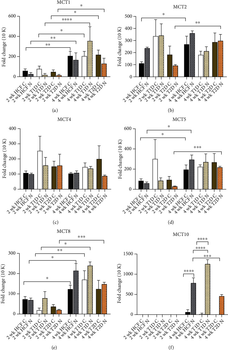
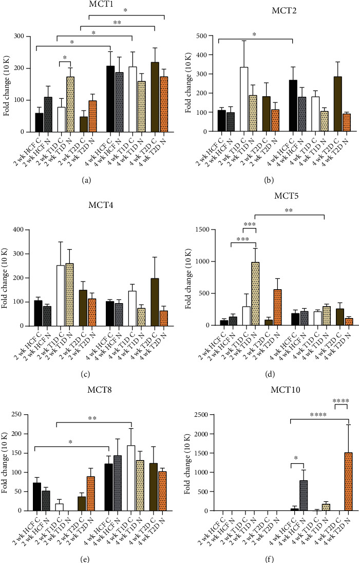
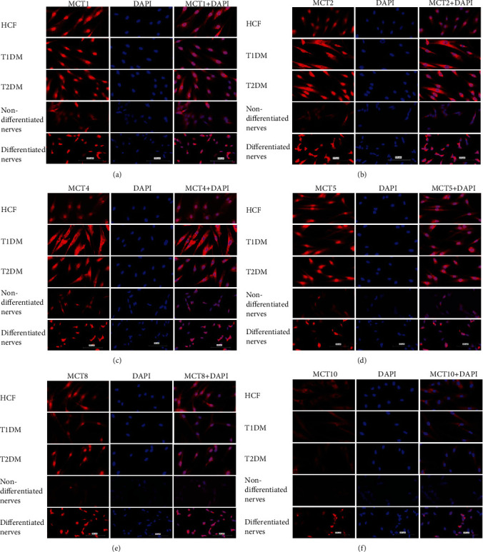
Diabetes mellitus (DM) is a group of metabolic diseases that is known to cause structural and functional ocular complications. In the human cornea, DM-related complications affect the epithelium, stroma, and nerves. Monocarboxylate transporters (MCTs) are a family of proton-linked plasma membrane transporters that carry monocarboxylates across plasma membranes. In the context of corneal health and disease, their role, presence, and function are largely undetermined and solely focused on the most common MCT isoforms, 1 through 4. In this study, we investigated the regulation of MCT1, 2, 4, 5, 8, and 10, in corneal DM, using established 3D self-assembled extracellular matrix (ECM) in vitro models. Primary stromal corneal fibroblasts were isolated from healthy (HCFs), type I (T1DMs), and type II (T2DMs) DM donors. Monoculture 3D constructs were created by stimulating stromal cells on transwells with stable vitamin C for two or four weeks. Coculture 3D constructs were created by adding SH-SY5Y neurons at two different densities, 12 k and 500 k, on top of the monocultures. Our data showed significant upregulation of MCT1 at 4 weeks for HCF, T1DM, and T2DM monocultures, as well as the 500 k nerve cocultures. MCT8 was significantly upregulated in HCF and T1DM monocultures and all of the 500 k nerve cocultures. Further, MCT10 was only expressed at 4 weeks for all cocultures and was limited to HCFs and T1DMs in monocultures. Immunofluorescence analysis showed cytoplasmic MCT expression for all cell types and significant downregulation of both MCT2 and MCT4 in HCFs, when compared to T1DMs and T2DMs. Herein, we reveal the existence and modulation of MCTs in the human diabetic cornea in vitro. Changes appeared dependent on neuronal density, suggesting that MCTs are very likely critical to the neuronal defects observed in diabetic keratopathy/neuropathy. Further studies are warranted in order to fully delineate the role of MCTs in corneal diabetes.


 分享
分享

 求助内容:
求助内容: 应助结果提醒方式:
 扫码关注我们
