Cheolwon Jang, Byung-Kyu Cho, Sung Hwan Hwang, Hyung Jin Shin, Sang Hoon Yoon
{"title":"Leptomeningeal Spread at the Diagnosis of Glioblastoma Multiforme: A Case Report and Literature Review.","authors":"Cheolwon Jang, Byung-Kyu Cho, Sung Hwan Hwang, Hyung Jin Shin, Sang Hoon Yoon","doi":"10.14791/btrt.2022.0013","DOIUrl":null,"url":null,"abstract":"<p><p>Approximately two-thirds of glioblastoma (GBM) patients progress to leptomeningeal spread (LMS) within two years. While 90% of LMS cases are diagnosed during the progression and/or recurrence of GBM (defined as secondary LMS), LMS presentation at the time of GBM diagnosis (defined as primary LMS) is very rare. <sup>18</sup>F-fluorodeoxy glucose positron emission tomography computed tomography (<sup>18</sup>F-FDG PET/CT) study helps to diagnose the multifocal spread of the malignant primary brain tumor. Our patient was a 31-year-old man with a tumorous lesion located in the right temporal lobe, a wide area of the leptomeninges, and spinal cord (thoracic 5/6, and lumbar 1 level) involvement as a concurrent manifestation. After the removal of the right temporal tumor, the clinical status progressed rapidly, showing signs of increased intracranial pressure and hydrocephalus caused by LMS. He underwent a ventriculoperitoneal shunt a week after craniotomy. During management, progression of cord compression, paraplegia, bone marrow suppression related to radiochemotherapy, intercurrent infections, and persistent ascites due to peritoneal metastasis of the LMS through the shunt system was observed. The patient finally succumbed to the disease nine months after the diagnosis of simultaneous GBM and LMS. The overall survival of primary LMS with GBM in our case was nine months, which is shorter than that of secondary LMS with GBM. The survival period after the diagnosis of LMS did not seem to be significantly different between primary and secondary LMS. To determine the prognostic effect and difference between primary and secondary LMS, further cooperative studies with large-volume data analysis are warranted.</p>","PeriodicalId":72453,"journal":{"name":"Brain tumor research and treatment","volume":"10 3","pages":"183-189"},"PeriodicalIF":0.0000,"publicationDate":"2022-07-01","publicationTypes":"Journal Article","fieldsOfStudy":null,"isOpenAccess":false,"openAccessPdf":"https://ftp.ncbi.nlm.nih.gov/pub/pmc/oa_pdf/1d/05/btrt-10-183.PMC9353161.pdf","citationCount":"0","resultStr":null,"platform":"Semanticscholar","paperid":null,"PeriodicalName":"Brain tumor research and treatment","FirstCategoryId":"1085","ListUrlMain":"https://doi.org/10.14791/btrt.2022.0013","RegionNum":0,"RegionCategory":null,"ArticlePicture":[],"TitleCN":null,"AbstractTextCN":null,"PMCID":null,"EPubDate":"","PubModel":"","JCR":"","JCRName":"","Score":null,"Total":0}
引用次数: 0
Abstract
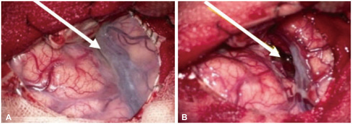
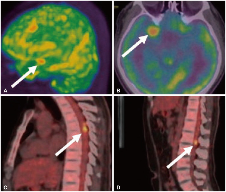
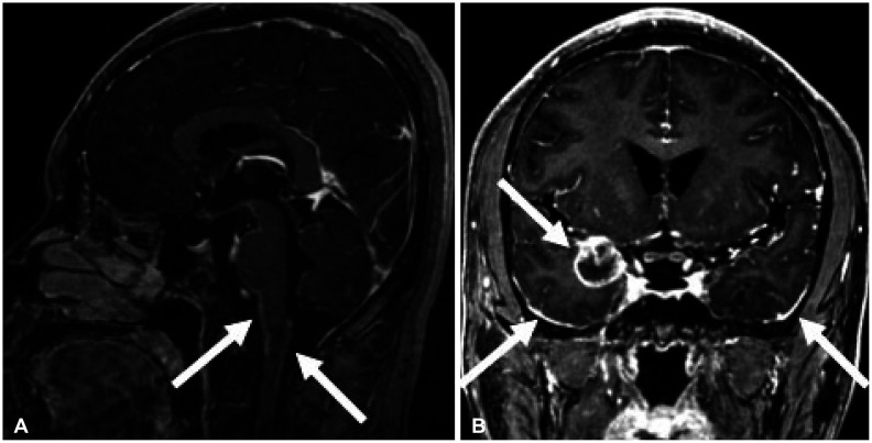
Approximately two-thirds of glioblastoma (GBM) patients progress to leptomeningeal spread (LMS) within two years. While 90% of LMS cases are diagnosed during the progression and/or recurrence of GBM (defined as secondary LMS), LMS presentation at the time of GBM diagnosis (defined as primary LMS) is very rare. 18F-fluorodeoxy glucose positron emission tomography computed tomography (18F-FDG PET/CT) study helps to diagnose the multifocal spread of the malignant primary brain tumor. Our patient was a 31-year-old man with a tumorous lesion located in the right temporal lobe, a wide area of the leptomeninges, and spinal cord (thoracic 5/6, and lumbar 1 level) involvement as a concurrent manifestation. After the removal of the right temporal tumor, the clinical status progressed rapidly, showing signs of increased intracranial pressure and hydrocephalus caused by LMS. He underwent a ventriculoperitoneal shunt a week after craniotomy. During management, progression of cord compression, paraplegia, bone marrow suppression related to radiochemotherapy, intercurrent infections, and persistent ascites due to peritoneal metastasis of the LMS through the shunt system was observed. The patient finally succumbed to the disease nine months after the diagnosis of simultaneous GBM and LMS. The overall survival of primary LMS with GBM in our case was nine months, which is shorter than that of secondary LMS with GBM. The survival period after the diagnosis of LMS did not seem to be significantly different between primary and secondary LMS. To determine the prognostic effect and difference between primary and secondary LMS, further cooperative studies with large-volume data analysis are warranted.


 分享
分享

 求助内容:
求助内容: 应助结果提醒方式:
 扫码关注我们
