{"title":"Spheno-Orbital Plasmacytoma as an Initial Presentation of Multiple Myeloma.","authors":"Sangjun Ahn, Seung Heon Cha, Won Ho Cho","doi":"10.14791/btrt.2022.0026","DOIUrl":null,"url":null,"abstract":"<p><p>Intracranial plasmacytoma is a rare neoplasm and a subtype of malignant plasma cell tumor. Most patients with plasma cell tumors are diagnosed with multiple myeloma, but 5%-10% of patients are not. This report includes descriptions of radiologic and clinical findings in a patient with intracranial plasmacytoma. Intracranial extra-axial plasmacytomas can be easily misdiagnosed as meningioma in radiologic and clinical findings. A 69-year-old woman presented with exophthalmos and diplopia, and MRI indicated meningioma. Thus, she underwent gross total resection, and her pathologic diagnosis was plasmacytoma. Exophthalmos and diplopia were fully recovered. She was finally diagnosed with multiple myeloma based on systemic evaluation and treated with targeted chemotherapy. MRI conducted at 3 months after surgery showed no local recurrence or remnant tumor. Although intracranial plasmacytomas are difficult to distinguish from meningiomas in preoperative evaluation, gross total resection is recommended for the same purposes as meningiomas. If the pathologic diagnosis is a plasmacytoma, it is essential to have a systemic evaluation for multiple myeloma.</p>","PeriodicalId":72453,"journal":{"name":"Brain tumor research and treatment","volume":"10 4","pages":"270-274"},"PeriodicalIF":0.0000,"publicationDate":"2022-10-01","publicationTypes":"Journal Article","fieldsOfStudy":null,"isOpenAccess":false,"openAccessPdf":"https://ftp.ncbi.nlm.nih.gov/pub/pmc/oa_pdf/97/b4/btrt-10-270.PMC9650124.pdf","citationCount":"0","resultStr":null,"platform":"Semanticscholar","paperid":null,"PeriodicalName":"Brain tumor research and treatment","FirstCategoryId":"1085","ListUrlMain":"https://doi.org/10.14791/btrt.2022.0026","RegionNum":0,"RegionCategory":null,"ArticlePicture":[],"TitleCN":null,"AbstractTextCN":null,"PMCID":null,"EPubDate":"","PubModel":"","JCR":"","JCRName":"","Score":null,"Total":0}
引用次数: 0
Abstract
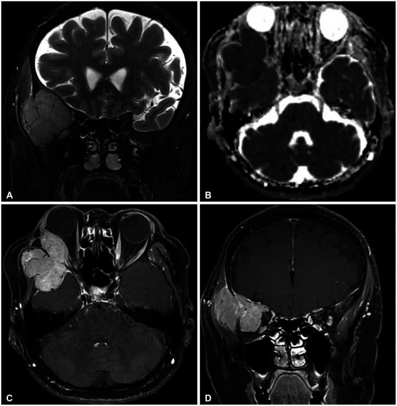
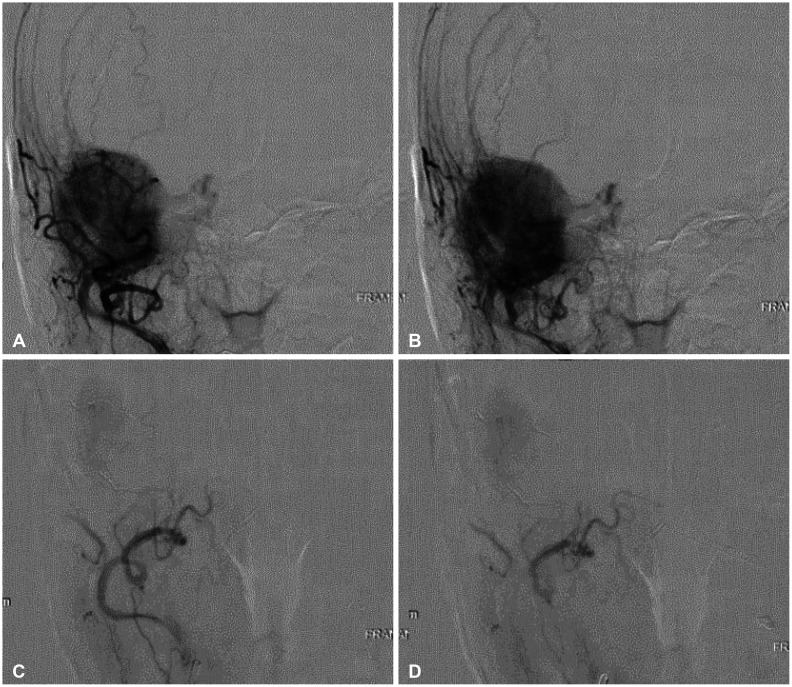
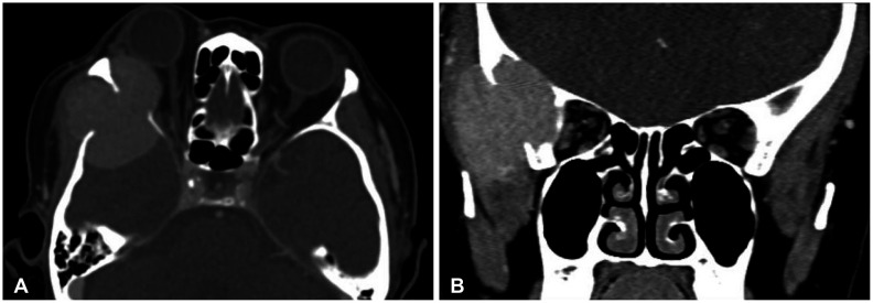
Intracranial plasmacytoma is a rare neoplasm and a subtype of malignant plasma cell tumor. Most patients with plasma cell tumors are diagnosed with multiple myeloma, but 5%-10% of patients are not. This report includes descriptions of radiologic and clinical findings in a patient with intracranial plasmacytoma. Intracranial extra-axial plasmacytomas can be easily misdiagnosed as meningioma in radiologic and clinical findings. A 69-year-old woman presented with exophthalmos and diplopia, and MRI indicated meningioma. Thus, she underwent gross total resection, and her pathologic diagnosis was plasmacytoma. Exophthalmos and diplopia were fully recovered. She was finally diagnosed with multiple myeloma based on systemic evaluation and treated with targeted chemotherapy. MRI conducted at 3 months after surgery showed no local recurrence or remnant tumor. Although intracranial plasmacytomas are difficult to distinguish from meningiomas in preoperative evaluation, gross total resection is recommended for the same purposes as meningiomas. If the pathologic diagnosis is a plasmacytoma, it is essential to have a systemic evaluation for multiple myeloma.


 分享
分享

 求助内容:
求助内容: 应助结果提醒方式:
 扫码关注我们
