{"title":"Imaging features of β-catenin-activated hepatocellular adenoma with weak β-catenin activation: A rare case report.","authors":"Kiyoyuki Minamiguchi, Nagaaki Marugami, Tomoko Uchiyama, Hironori Kusano, Satoshi Yasuda, Masayuki Sho, Toshihiro Tanaka","doi":"10.1177/20584601221142241","DOIUrl":null,"url":null,"abstract":"<p><p>We report valuable imaging findings in a case of β-catenin-activated hepatocellular adenoma (β-HCA) with weak β-catenin activation. A 40 year-old female presented with a liver tumor in S8 that was incidentally detected on ultrasonography. The tumor showed marked enhancement and early venous drainage into the middle hepatic vein in the arterial phase of contrast-enhanced computed tomography (CT). The tumor revealed slight hypointensity in the hepatobiliary phase of gadolinium ethoxybenzyl diethylenetriamine pentaacetic acid-enhanced magnetic resonance imaging (EOB-MRI). Six months after detection, the tumor had increased in size and a biopsy indicated hepatocellular carcinoma. The tumor was resected and pathologically diagnosed as β-HCA with weak β-catenin activation such as exon 3 S45 mutation and exon 7/8 mutation. Marked enhancement in the arterial phase of CT and MRI is a characteristic finding of β-HCA with weak β-catenin activation. Furthermore, the degree of β-catenin activation might determine the signal intensity of β-HCA in the hepatobiliary phase of EOB-MRI.</p>","PeriodicalId":72063,"journal":{"name":"Acta radiologica open","volume":"11 11","pages":"20584601221142241"},"PeriodicalIF":1.0000,"publicationDate":"2022-11-23","publicationTypes":"Journal Article","fieldsOfStudy":null,"isOpenAccess":false,"openAccessPdf":"https://ftp.ncbi.nlm.nih.gov/pub/pmc/oa_pdf/89/21/10.1177_20584601221142241.PMC9693779.pdf","citationCount":"1","resultStr":null,"platform":"Semanticscholar","paperid":null,"PeriodicalName":"Acta radiologica open","FirstCategoryId":"1085","ListUrlMain":"https://doi.org/10.1177/20584601221142241","RegionNum":0,"RegionCategory":null,"ArticlePicture":[],"TitleCN":null,"AbstractTextCN":null,"PMCID":null,"EPubDate":"2022/11/1 0:00:00","PubModel":"eCollection","JCR":"Q4","JCRName":"RADIOLOGY, NUCLEAR MEDICINE & MEDICAL IMAGING","Score":null,"Total":0}
引用次数: 1
Abstract
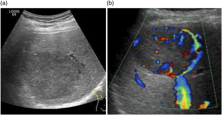
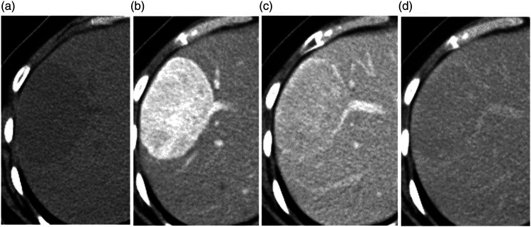
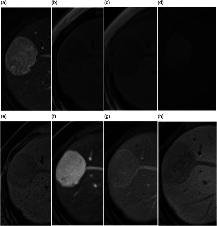
We report valuable imaging findings in a case of β-catenin-activated hepatocellular adenoma (β-HCA) with weak β-catenin activation. A 40 year-old female presented with a liver tumor in S8 that was incidentally detected on ultrasonography. The tumor showed marked enhancement and early venous drainage into the middle hepatic vein in the arterial phase of contrast-enhanced computed tomography (CT). The tumor revealed slight hypointensity in the hepatobiliary phase of gadolinium ethoxybenzyl diethylenetriamine pentaacetic acid-enhanced magnetic resonance imaging (EOB-MRI). Six months after detection, the tumor had increased in size and a biopsy indicated hepatocellular carcinoma. The tumor was resected and pathologically diagnosed as β-HCA with weak β-catenin activation such as exon 3 S45 mutation and exon 7/8 mutation. Marked enhancement in the arterial phase of CT and MRI is a characteristic finding of β-HCA with weak β-catenin activation. Furthermore, the degree of β-catenin activation might determine the signal intensity of β-HCA in the hepatobiliary phase of EOB-MRI.


 分享
分享

 求助内容:
求助内容: 应助结果提醒方式:
 扫码关注我们
