Gyu-Dong Jo, Ju-Hee Kang, Jo-Eun Kim, Won-Jin Yi, Min-Suk Heo, Sam-Sun Lee, Kyung-Hoe Huh
{"title":"Head and neck manifestations of fibrodysplasia ossificans progressiva: Clinical and imaging findings in 2 cases.","authors":"Gyu-Dong Jo, Ju-Hee Kang, Jo-Eun Kim, Won-Jin Yi, Min-Suk Heo, Sam-Sun Lee, Kyung-Hoe Huh","doi":"10.5624/isd.20230069","DOIUrl":null,"url":null,"abstract":"<p><p>Fibrodysplasia ossificans progressiva is a rare hereditary disorder characterized by progressive heterotopic ossification in muscle and connective tissue, with few reported cases affecting the head and neck region. Although plain radiographic findings and computed tomography features have been well documented, limited reports exist on magnetic resonance findings. This report presents 2 cases of fibrodysplasia ossificans progressiva, one with limited mouth opening due to heterotopic ossification of the lateral pterygoid muscle and the other with restricted neck movement due to heterotopic ossification of the platysma muscle. Clinical findings of restricted mouth opening or limited neck movement, along with radiological findings of associated heterotopic ossification, should prompt consideration of fibrodysplasia ossificans progressiva in the differential diagnosis. Dentists should be particularly vigilant with patients diagnosed with fibrodysplasia ossificans progressiva to avoid exposure to diagnostic biopsy and invasive dental procedures.</p>","PeriodicalId":51714,"journal":{"name":"Imaging Science in Dentistry","volume":"53 3","pages":"257-264"},"PeriodicalIF":2.1000,"publicationDate":"2023-09-01","publicationTypes":"Journal Article","fieldsOfStudy":null,"isOpenAccess":false,"openAccessPdf":"https://ftp.ncbi.nlm.nih.gov/pub/pmc/oa_pdf/46/a0/isd-53-257.PMC10548149.pdf","citationCount":"0","resultStr":null,"platform":"Semanticscholar","paperid":null,"PeriodicalName":"Imaging Science in Dentistry","FirstCategoryId":"1085","ListUrlMain":"https://doi.org/10.5624/isd.20230069","RegionNum":0,"RegionCategory":null,"ArticlePicture":[],"TitleCN":null,"AbstractTextCN":null,"PMCID":null,"EPubDate":"2023/6/20 0:00:00","PubModel":"Epub","JCR":"Q3","JCRName":"DENTISTRY, ORAL SURGERY & MEDICINE","Score":null,"Total":0}
引用次数: 0
Abstract
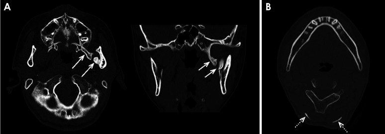
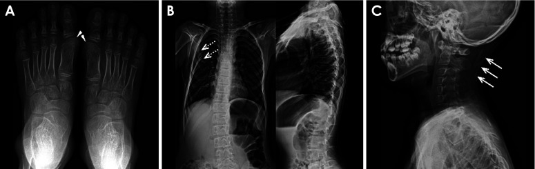
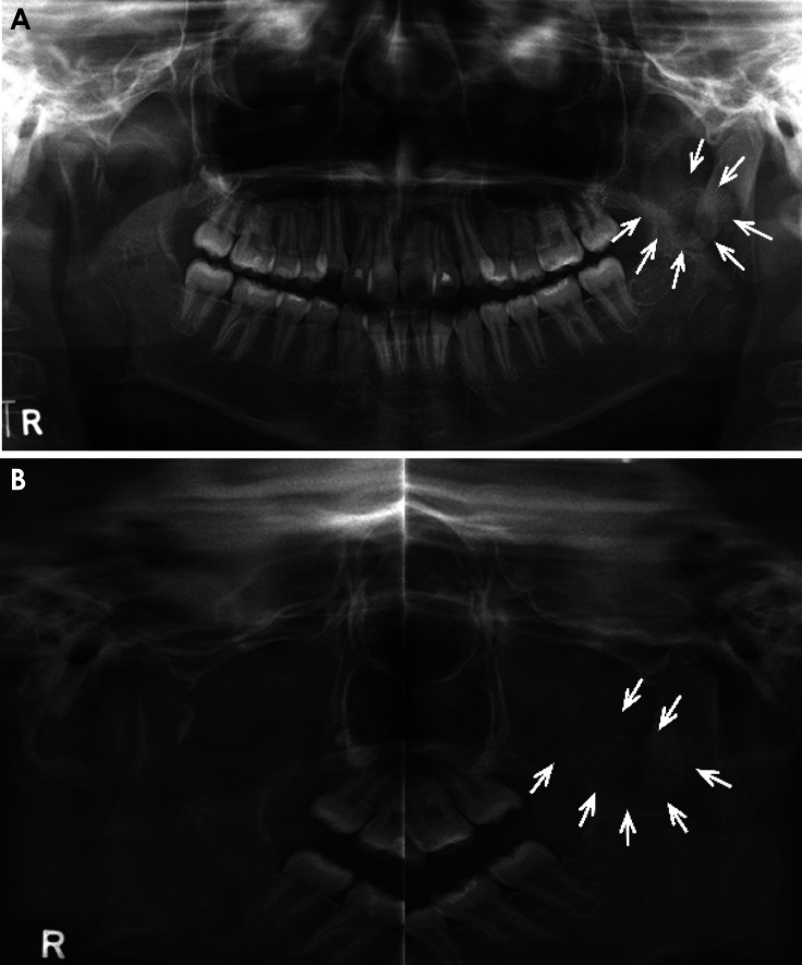
Fibrodysplasia ossificans progressiva is a rare hereditary disorder characterized by progressive heterotopic ossification in muscle and connective tissue, with few reported cases affecting the head and neck region. Although plain radiographic findings and computed tomography features have been well documented, limited reports exist on magnetic resonance findings. This report presents 2 cases of fibrodysplasia ossificans progressiva, one with limited mouth opening due to heterotopic ossification of the lateral pterygoid muscle and the other with restricted neck movement due to heterotopic ossification of the platysma muscle. Clinical findings of restricted mouth opening or limited neck movement, along with radiological findings of associated heterotopic ossification, should prompt consideration of fibrodysplasia ossificans progressiva in the differential diagnosis. Dentists should be particularly vigilant with patients diagnosed with fibrodysplasia ossificans progressiva to avoid exposure to diagnostic biopsy and invasive dental procedures.


 分享
分享
 求助内容:
求助内容: 应助结果提醒方式:
 扫码关注我们
