Jéssica Albuquerque M Silva, Bruno Hochhegger, Viviane Brandão Amorim, Gláucia Zanetti, Edson Marchiori
{"title":"Computed tomography aspects of thoracic metastases from osteosarcoma: pictorial essay.","authors":"Jéssica Albuquerque M Silva, Bruno Hochhegger, Viviane Brandão Amorim, Gláucia Zanetti, Edson Marchiori","doi":"10.1590/0100-3984.2022.0107-en","DOIUrl":null,"url":null,"abstract":"<p><p>Osteosarcoma is the most common primary bone tumor, with a higher incidence in the second decade of life, and it often leads to pulmonary metastases. The most common pattern seen on computed tomography is one of multiple well-defined nodules in the lung parenchyma, often with calcifications. Because of the variety of presentations of pulmonary metastases in osteosarcoma, including atypical forms, knowledge of the computed tomography aspects of these lesions is important for characterizing and evaluating the extent of the disease, as well as for distinguishing metastatic disease from other benign or malignant lung diseases. This essay discusses the main tomographic findings of pulmonary metastases from osteosarcoma.</p>","PeriodicalId":20842,"journal":{"name":"Radiologia Brasileira","volume":"56 4","pages":"215-219"},"PeriodicalIF":0.0000,"publicationDate":"2023-07-01","publicationTypes":"Journal Article","fieldsOfStudy":null,"isOpenAccess":false,"openAccessPdf":"https://www.ncbi.nlm.nih.gov/pmc/articles/PMC10567086/pdf/","citationCount":"0","resultStr":null,"platform":"Semanticscholar","paperid":null,"PeriodicalName":"Radiologia Brasileira","FirstCategoryId":"1085","ListUrlMain":"https://doi.org/10.1590/0100-3984.2022.0107-en","RegionNum":0,"RegionCategory":null,"ArticlePicture":[],"TitleCN":null,"AbstractTextCN":null,"PMCID":null,"EPubDate":"","PubModel":"","JCR":"Q3","JCRName":"Medicine","Score":null,"Total":0}
引用次数: 0
Abstract
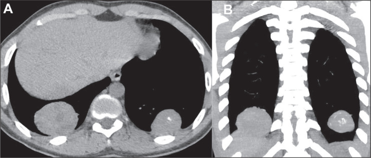
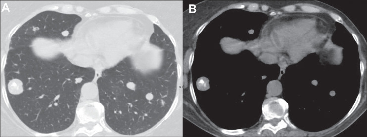
Osteosarcoma is the most common primary bone tumor, with a higher incidence in the second decade of life, and it often leads to pulmonary metastases. The most common pattern seen on computed tomography is one of multiple well-defined nodules in the lung parenchyma, often with calcifications. Because of the variety of presentations of pulmonary metastases in osteosarcoma, including atypical forms, knowledge of the computed tomography aspects of these lesions is important for characterizing and evaluating the extent of the disease, as well as for distinguishing metastatic disease from other benign or malignant lung diseases. This essay discusses the main tomographic findings of pulmonary metastases from osteosarcoma.


 分享
分享
 求助内容:
求助内容: 应助结果提醒方式:
 扫码关注我们
