{"title":"Commissure leaflet prolapse closely mimics anterior mitral leaflet perforation in 2-D image of transesophageal echocardiography.","authors":"Kazuto Miyata, Sayaka Shigematsu, Naoki Miyayama","doi":"10.1186/s40981-023-00659-z","DOIUrl":null,"url":null,"abstract":"<p><strong>Background: </strong>Precise diagnosis of mitral valve regurgitation is challenging, particularly for distinguishing between commissure leaflet prolapse and anterior leaflet perforation, based exclusively on 2-dimensional (2-D) imaging by transesophageal echocardiography. CASE 1: Two mitral regurgitation jets suggesting anterior leaflet perforation, but no regurgitation orifices, were observed in the mid esophageal (ME) 4-chamber view. Multiple 2-D and 3-dimensional (3-D) images revealed prolapse of the anterior (A3) leaflet and posterior commissure, not anterior leaflet perforation. CASE 2: A regurgitation jet suggesting an anterior leaflet prolapse with a regurgitation orifice was observed in ME long-axis view. Multiple 2-D and 3-D images showed only anterior commissure prolapse, but no signs of anterior leaflet perforation.</p><p><strong>Conclusions: </strong>A regurgitant jet caused by commissure leaflet prolapse closely resembles anterior leaflet perforation in 2-D imaging. Careful evaluation of multiple 2-D and 3-D images, as well as of the regurgitation orifices, is crucially important for making an accurate diagnosis.</p>","PeriodicalId":14635,"journal":{"name":"JA Clinical Reports","volume":"9 1","pages":"67"},"PeriodicalIF":1.0000,"publicationDate":"2023-10-16","publicationTypes":"Journal Article","fieldsOfStudy":null,"isOpenAccess":false,"openAccessPdf":"https://www.ncbi.nlm.nih.gov/pmc/articles/PMC10579197/pdf/","citationCount":"0","resultStr":null,"platform":"Semanticscholar","paperid":null,"PeriodicalName":"JA Clinical Reports","FirstCategoryId":"1085","ListUrlMain":"https://doi.org/10.1186/s40981-023-00659-z","RegionNum":0,"RegionCategory":null,"ArticlePicture":[],"TitleCN":null,"AbstractTextCN":null,"PMCID":null,"EPubDate":"","PubModel":"","JCR":"Q3","JCRName":"ANESTHESIOLOGY","Score":null,"Total":0}
引用次数: 0
Abstract
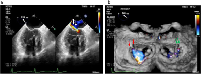
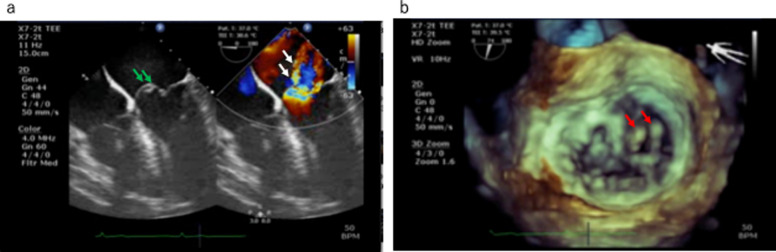
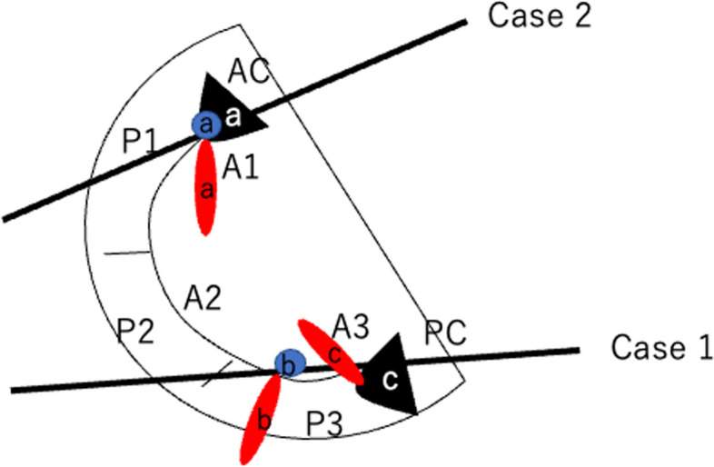
Background: Precise diagnosis of mitral valve regurgitation is challenging, particularly for distinguishing between commissure leaflet prolapse and anterior leaflet perforation, based exclusively on 2-dimensional (2-D) imaging by transesophageal echocardiography. CASE 1: Two mitral regurgitation jets suggesting anterior leaflet perforation, but no regurgitation orifices, were observed in the mid esophageal (ME) 4-chamber view. Multiple 2-D and 3-dimensional (3-D) images revealed prolapse of the anterior (A3) leaflet and posterior commissure, not anterior leaflet perforation. CASE 2: A regurgitation jet suggesting an anterior leaflet prolapse with a regurgitation orifice was observed in ME long-axis view. Multiple 2-D and 3-D images showed only anterior commissure prolapse, but no signs of anterior leaflet perforation.
Conclusions: A regurgitant jet caused by commissure leaflet prolapse closely resembles anterior leaflet perforation in 2-D imaging. Careful evaluation of multiple 2-D and 3-D images, as well as of the regurgitation orifices, is crucially important for making an accurate diagnosis.
期刊介绍:
JA Clinical Reports is a companion journal to the Journal of Anesthesia (JA), the official journal of the Japanese Society of Anesthesiologists (JSA). This journal is an open access, peer-reviewed, online journal related to clinical anesthesia practices such as anesthesia management, pain management and intensive care. Case reports are very important articles from the viewpoint of education and the cultivation of scientific thinking in the field of anesthesia. However, submissions of anesthesia research and clinical reports from Japan are notably decreasing in major anesthesia journals. Therefore, the JSA has decided to launch a new journal, JA Clinical Reports, to encourage JSA members, particularly junior Japanese anesthesiologists, to publish papers in English language.


 分享
分享
 求助内容:
求助内容: 应助结果提醒方式:
 扫码关注我们
