Yena Kwon, Byeong-Seon An, Yeon-Ju Shin, Cheol-Woong Yang
{"title":"Method of Ga removal from a specimen on a microelectromechanical system-based chip for in-situ transmission electron microscopy","authors":"Yena Kwon, Byeong-Seon An, Yeon-Ju Shin, Cheol-Woong Yang","doi":"10.1186/s42649-020-00043-6","DOIUrl":null,"url":null,"abstract":"<p>In-situ transmission electron microscopy (TEM) holders that employ a chip-type specimen stage have been widely utilized in recent years. The specimen on the microelectromechanical system (MEMS)-based chip is commonly prepared by focused ion beam (FIB) milling and ex-situ lift-out (EXLO). However, the FIB-milled thin-foil specimens are inevitably contaminated with Ga<sup>+</sup> ions. When these specimens are heated for real time observation, the Ga<sup>+</sup> ions influence the reaction or aggregate in the protection layer. An effective method of removing the Ga residue by Ar<sup>+</sup> ion milling within FIB system was explored in this study. However, the Ga residue remained in the thin-foil specimen that was extracted by EXLO from the trench after the conduct of Ar<sup>+</sup> ion milling. To address this drawback, the thin-foil specimen was attached to an FIB lift-out grid, subjected to Ar<sup>+</sup> ion milling, and subsequently transferred to an MEMS-based chip by EXLO. The removal of the Ga residue was confirmed by energy dispersive spectroscopy.</p>","PeriodicalId":470,"journal":{"name":"Applied Microscopy","volume":"50 1","pages":""},"PeriodicalIF":0.0000,"publicationDate":"2020-10-14","publicationTypes":"Journal Article","fieldsOfStudy":null,"isOpenAccess":false,"openAccessPdf":"https://sci-hub-pdf.com/10.1186/s42649-020-00043-6","citationCount":"1","resultStr":null,"platform":"Semanticscholar","paperid":null,"PeriodicalName":"Applied Microscopy","FirstCategoryId":"1085","ListUrlMain":"https://link.springer.com/article/10.1186/s42649-020-00043-6","RegionNum":0,"RegionCategory":null,"ArticlePicture":[],"TitleCN":null,"AbstractTextCN":null,"PMCID":null,"EPubDate":"","PubModel":"","JCR":"Q3","JCRName":"Immunology and Microbiology","Score":null,"Total":0}
引用次数: 1
Abstract
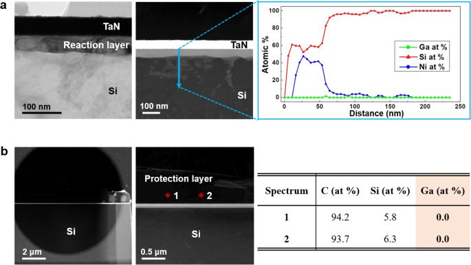
In-situ transmission electron microscopy (TEM) holders that employ a chip-type specimen stage have been widely utilized in recent years. The specimen on the microelectromechanical system (MEMS)-based chip is commonly prepared by focused ion beam (FIB) milling and ex-situ lift-out (EXLO). However, the FIB-milled thin-foil specimens are inevitably contaminated with Ga+ ions. When these specimens are heated for real time observation, the Ga+ ions influence the reaction or aggregate in the protection layer. An effective method of removing the Ga residue by Ar+ ion milling within FIB system was explored in this study. However, the Ga residue remained in the thin-foil specimen that was extracted by EXLO from the trench after the conduct of Ar+ ion milling. To address this drawback, the thin-foil specimen was attached to an FIB lift-out grid, subjected to Ar+ ion milling, and subsequently transferred to an MEMS-based chip by EXLO. The removal of the Ga residue was confirmed by energy dispersive spectroscopy.
Applied MicroscopyImmunology and Microbiology-Applied Microbiology and Biotechnology
CiteScore
3.40
自引率
0.00%
发文量
10
审稿时长
10 weeks
期刊介绍:
Applied Microscopy is a peer-reviewed journal sponsored by the Korean Society of Microscopy. The journal covers all the interdisciplinary fields of technological developments in new microscopy methods and instrumentation and their applications to biological or materials science for determining structure and chemistry. ISSN: 22875123, 22874445.


 分享
分享
 求助内容:
求助内容: 应助结果提醒方式:
 扫码关注我们
